|


|
synonyms:
Olecranon Stress Fracture ICD-10
A- initial encounter for fracture
D- subsequent encounter for fracture with routine healing
G- subsequent encounter for fracture with delayed healing
K- subsequent encounter for fracture with nonunion
P- subsequent encounter for fracture with malunion
S- sequela
Olecranon Stress Fracture ICD-9
- 733.95 Stress fracture of other bone
Olecranon Stress Fracture Etiology / Epidemiology / Natural History
- Generally associated with throwning sports, baseball, javelin (Waris W. Acta Chir Scand 1946;93:563).
- May be related to triceps overload (Kvidera DJ, Orthop Rev 1983;12:113).
Olecranon Stress Fracture Anatomy
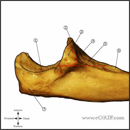
- Olecranon stress fractures may occur through the physis in adolescent athletes. (Lowery WD, JSES 1995;4:143), (Maffulti N, JBJS Br 1992;74B:305).
- Oblique olecranon stress fractures are reported in throwing athletes with a closed physis. (Nuber GW, Clin Orthop 1992;278:58), (Suzuki K, JSES 1997;6:492).
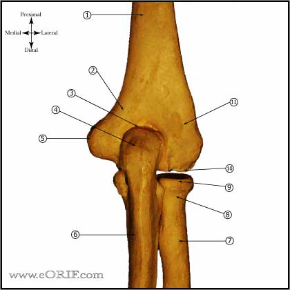
- see also Elbow Anatomy.
Olecranon Stress Fracture Clinical Evaluation
- Complain of elbow pain, more commonly in the acceleration phase of throwing.
- Tenderness over the olecranon. May also have tenderness over the anterior band of the MCL.
Olecranon Stress Fracture Xray / Diagnositc Tests
- A/P, Lateral and oblique elbow xrays. Generally demonstrate olecranon stress fracture, with sclerotic changes. If no fracture is seen, but suspicion is high consider MRI or CT.
- MRI arthrogram: study of choice for elbow MCL injury, but also demonstrates olecranon stress fractures well. Evaluate for T sign of ulnar avulsion, humeral MCL avulsion or midsubstance tearing. Normal MCL appears as a thin band of low intensity along the medial elbow. Tearing is indicated by signal attenuation or absence. (Timmerman LA, AJSM 1994;22:26), (Munshi M, Radiology 2004;231:797), (Schwartz ML, Radiology 1995;197: 297).
82. Sisto DJ, Jobe FW, Moynes DR, et al: An el
- CT:
Olecranon Stress Fracture Classification / Treatment
- Treatment: rest and temporary splinting. Consider electrical stimulation. ORIF indicated after failure of nonoperative treatment. (Ahmad CS, Clin Sports Med 2004;23:665) (Rettig AC, AJSM 2006;34:653). Early ORIF may be indicated oblique olecranon stress fractures with sclerotic edges (Suzuki K, JSES 1997;6:492).
- Olecranon ORIF: sclerotic areas are often difficult to drill and place screws through. Consider taping before screw placement if difficult to drill. Consider bone grafting or using bone graft substitutes.
Olecranon Stress Fracture Associated Injuries / Differential Diagnosis
Olecranon Stress Fracture Complications
Olecranon Stress Fracture Follow-up Care
- Post-op: Place in posterior splint in 50-70º of flexion. Encourage finger ROM.
- 10-14 Days: Wound check, remove staples. Evaluate reduction on xrays. Place in long-arm cast or removable posterior splint depending on fracture type and strength of fixation.
- 6 Weeks: Wound check, evaluate xrays for callus formation. Place in removable posterior splint and begin elbow ROM exercises.
- 3 Months: Evaluate xrays for fracture union. Begin gentle sport specific exercises depending on fracture union. Consider bone stimulator if union not evident on xrays.
- 6 Months: Evaluate xrays. Return to sport.
- 1Yr: Evaluate xrays. Evaluate xrays using Elbow Outcome Measures. May consider Hardware Removal if hardware is painful.
Olecranon Stress Fracture Review References
|


