 |
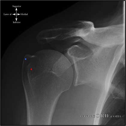
True AP Shoulder in neutral rotation (taken in the plane of the scapula) (Grashey view)
Blue dot = Greater Tuberosity Position: Patient erect, turned 30-35° toward the side being xrayed |
 |
AP Shoulder in External rotation (taken in the plane of the scapula)
Blue dot = Greater Tuberosity
Position: Patient erect, turned 30-35° toward the side being xrayed; arm maximally externally rotated |
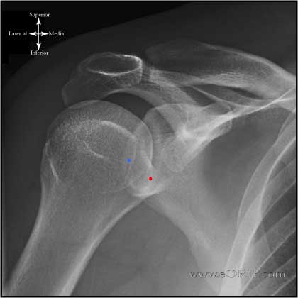 |
AP Shoulder in Internal rotation (taken in the plane of the scapula)
Blue dot = Greater Tuberosity
Position: Patient erect, turned 30-35° toward the side being xrayed; arm maximally internally rotated Beam: aimed perpendicular to plate |
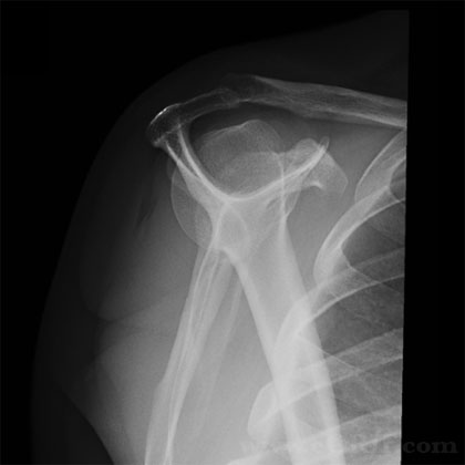 |
Scapular Y Shoulder Xray
Position: Erect with anterior aspect of affected shoulder against x-ray plate and rotating other shoulder out 40 deg°. Beam: aimed from posteriorly along scapular spine |
 |
Axillary view Shoulder Xray
Position: Patient seated at side of radiographic table with the arm abducted and axilla over the cassette. Beam:angle 5°-10° toward the elbow, central beam directed at the shoulder joint. Many alternative postions for similar xray, can be supine etc. |
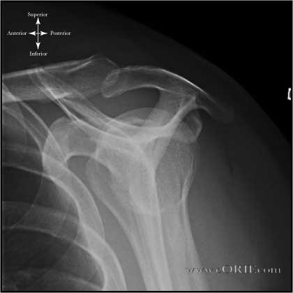 |
Supraspinatus Outlet view Shoulder Xray
Position: Erect with anterior aspect of affected shoulder against x-ray plate and rotating other shoulder out 40 deg°. Beam: aimed from posteriorly along scapular spine but with the beam aimed with 10° caudal tilt |
 |
Zanca View Shoulder Xray
Position: Erected with cassette behind shoulder. Beam:Xray beam aimed at the AC joint in 10° to 15° cephalic tilt. Xray penetration should be 1/2 normal to avoid overpenetration of AC joint. |
 |
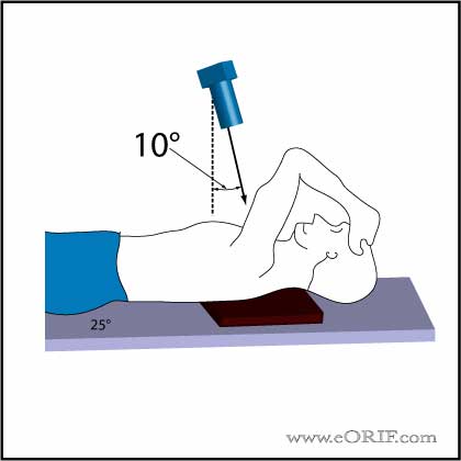
West Point Axillary View Shoulder Xray
Postion:Patient prone with affected shoulder resting on a pad @8cm for the table top. Casette positioned against the superior apsect of the shoulder. Beam: aimed 25° from horizontal (to tables surface) and 25° medially (to patients midline).
(Rokous JR, CORR 1972;82:84) |
 |
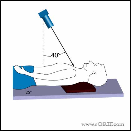
Stryker Notch View Shoulder Xray
Position: Patient supine with cassette posterior to the shoulder. The hand placed on top of the head. The elbow should point straight upward. Beam directed 10° superiorly/toward the head, centered over the coracoid process.
(Hall RH, JBJS 1959;41-A:489-94) |
 |
Serendipity View Shoulder Xray
Postion: supine with cassette under upper chest |
|
Bennett's View (modified)Shoulder Xray
|
|
 |
Bennett's view Shoulder Xray
|
 |
Acromiohumeral Interval
|
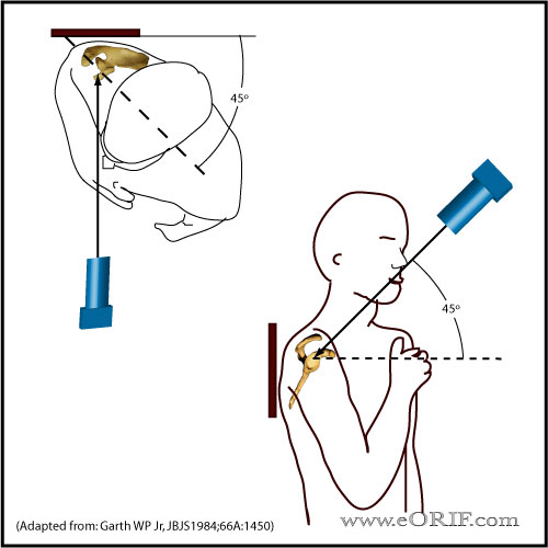 |
Garth View (apical oblique) Shoulder Xray
Postion: Seated with shoulder adjacent to cassette and arm adducted and internally rotated (place hand over heart). Chest rotated 45º Beam: beam perpendicular to the anterior-inferior glenoid rim and posterior-superior humeral head. (45º to the coronal plane and 45º caudally). Rollover for example rendition. Garth WP Jr, JBJS1984;66A:1450 |
