|


|
synonyms: Hill-Sachs lesion, reverse Hill-Sachs, compression fracture of humeral head,
Hill-Sachs Lesion ICD-10
A- initial encounter for closed fracture
B- initial encounter for open fracture
D- subsequent encounter for fracture with routine healing
G- subsequent encounter for fracture with delayed healing
K- subsequent encounter for fracture with nonunion
P- subsequent encounter for fracture with malunion
S- sequela
Hill-Sachs Lesion ICD-9
- 812.00 (fracture of humerus, upper end, unspecified)
- 736.89 other deformity of arm or leg , no elsewhere classified
- 733.90 other deformity of bone and cartilage, unspecified
Hill-Sachs Lesion Etiology / Epidemiology / Natural History
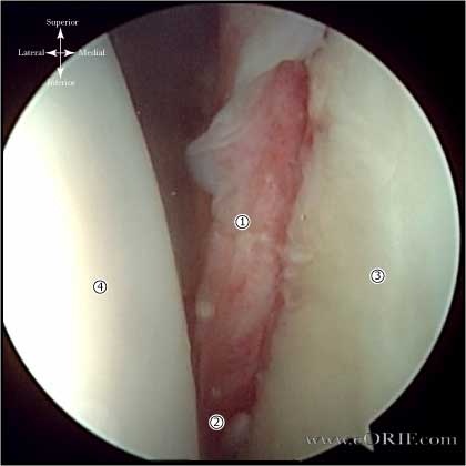
- Hill-Sachs Lesion = Impression fracture of the posterolateral humeral head; produced by contact with the anteroinferior glenoid when dislocated.
- Present in 90% of anterior dislocations and 25% of anterior subluxations. (Calandra JJ, Arthroscopy 1989;5:254).
- 84% of patients with recurrent instability have Hill-Sachs lesions (Boileau P, JBJS 2006;88A:1755).
- Reverse Hill-Sachs Lesion = impression fracture in the anterolateral humera head; related to posterior instability
- Engaging Hill-Sachs lesion = a defect in the humeral head large enough that the edge drops over the glenoid rim when the arm is externally rotated.
Hill-Sachs Lesion Anatomy
Hill-Sachs Lesion Clinical Evaluation
Hill-Sachs Lesion Xray / Diagnositc Tests
- Hill-Sachs lesion:best demonstrated on plain AP radiograph in internal rotation or Stryker notch view. Reverse Hill-Sachs lesion best seen on Grashey view with arm in external rotation.
- A/P and Lateral view in the plane of the scapula, and axillary view. Generally normal.
- West Point view: patient prone with arm in 90° abduction and neutral rotation. Xray beam is directed 25° posterior to the horizontal plane and 25° medial to the vertical plane. Useful for evaluating the anterior glenoid rim / bony bankart lesions.
- Garth (apical oblique) view: provides view of glenoid bone loss and Hill-Sachs lesions.
- CT scan is best to evaluate bony anatomy and should be considered for the recurrent dislocator suspected of having a large Hill-Sachs or bony Bankart lesion.
- MRI arthrogram (gadolinium): the anterior and posterior labrum are best seen on axial images and appear as dark triangular structures. Bankart lesions appear as a loss of the normal triangular shape or contrast material may extend between the labrum and glenoid( acute injuries.) Chronic injuries may scar down to the glenoid and be difficult to see by MRI.
Hill-Sachs Lesion Classification / Treatment
- Grading (Franceshi R, AJSM 2008;36:1330)
grade I (cartilaginous),
grade II (superficial bony scuffing)
grade III (hatchet fracture)
- Hill-Sachs lesion Classification (Calandra JJ, Arthroscopy. 1989;5(4):254): Grade 1: articular surface defect down to the subchondral bone, Grade 2: articular surface defect including the subchondral bone, Grade 3: a large defect in the subchondral bone
- Most Hill-Sachs lesions are small and do not require treatment.
- Each lesion should be evaluated during surgery. Lesions found to be engaging in a normal ROM require treatment.
- Lesions representing >30% of the articular surface (determined on pre-op CT) generally require treatment.
- Treatment options: humeral head allograft (Gerber C, JBJS 1996;78A:376), Remplissage (Wolf EM, Arthrosopy 2004;20(suppl1):e14), Humeral rotation osteotomy (Weber BG, JBJS 1984;66A:1443), Hemiarthroplasty / Total shoulder arthroplasty for patients >50y/o (Flatow E, JSES 1993;2:2).
Hill-Sachs Lesion Associated Injuries / Differential Diagnosis
- Anterior Glenohumeral Instability
- Hills Sachs lesion
- Posterior Glenohumeral Instability
- Internal Impingement
- RTC Tear
- Posterior Capular contracture
- SLAP Lesion
- Subacromial Impingement
- HAGL Lesion
- Suprascapular nerve entrapment(Cummins CA JBJS-AM 2000;82:415-424).
- Quadrilateral space syndrome(Cahill BR J Hand Surg-AM 1983;8:65-69).
- Early osteoarthritis
- Tumor
Hill-Sachs Lesion Complications
- Recurrent instability
- Pain
- Hardware failure / Anchor pull-out
- Infections
- Stiffness
- CRPS
- Nerve injury: Axillary nerve, Brachial plexus
- Fluid Extravasation:
- Chondrolysis: though to be related to heat from electo cautery or radiofrequency probes used during capsular release or capsular shrinkage.
- Hematoma
- Chondral Injury / arthritis
- Instrument failure
- Weakness
Hill-Sachs Lesion Follow-up Care
Hill-Sachs Lesion Review References
- Lynch JR, JSES 2009;18:317
|


