|


|
synonyms:olecranon fracture ORIF, elbow fracture ORIF
Olecranon Fracture ORIF CPT
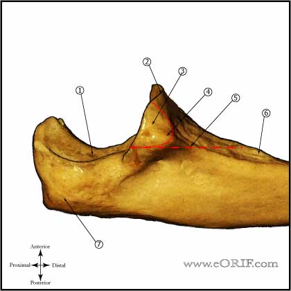
Olecranon Fracture ORIF Anatomy
Olecranon Fracture ORIF Indications
- Displaced olecranon fracture
Olecranon Fracture ORIF Contraindications
- Active infection
- Medically unstable for sugery
Olecranon Fracture ORIF Alternatives
- Non-operative treatment
- External Fixation
Olecranon Fracture ORIF Pre-op Planning
- Consider using pre-countoured plates
- For fractures with comminution, radiocapitellar or transolecranon instability, articular marginal impaction should be treated with Plate fixation.
- Consider a tension band wire construct for simple fracture patterns without comminution.
- Consider excision and triceps advancement in elderly, low-demand patients with small unreconstructable fracture patterns without associated elbow instability.
Olecranon Fracture ORIF Technique
- Sign operative site.
- Pre-operative antibiotics, +/- regional block.
- General endotracheal anesthesia
- position. All bony prominences well padded.
- Examination under anesthesia.
- Prep and drape in standard sterile fashion.
- Midline posterior incision curved around the olecranon.
- Fracture opened and hematoma debrided.
- Anatomic reduction with large tenaculum clamp. Consider drill hole in dorsal ulna to provide anchor point for clamp.
- Place two parallel 0.062 or 0.045 k-wires obliquely from proximal ulna, across fracture site and into the anterior ulnar cortex. Back them up 5mm to allow later impaction.
- Place 2.5mm drill hole transversely across triangular ulna diaphysis distal to the fracture and anterior enough to be bicortical.
- Place one 18-gauge wire or two 22-gauge wires (using two tranvserse distal drill holes) in figure-8 fashion through transverse hole and under the triceps insertion and k-wires proximally.
- Tighed the wires both medially and laterally. Trim twisted ends and bend into the adjacent soft tissue.
- Bend k-wires and cut excess.
- Incise triceps and impact bent ends into the proximal ulna with bone tamp.
- Irrigate.
- Close in layers.
Olecranon Fracture ORIF Complications
Olecranon Fracture ORIF Follw-up care
- Post-op: Place in posterior splint in 50-70º of flexion. Encourage finger ROM.
- 10-14 Days: Wound check, remove staples. Evaluate reduction on xrays. Place in long-arm cast or removable posterior splint depending on fracture type and strength of fixation.
- 6 Weeks: Wound check, evaluate xrays for callus formation. Place in removable posterior splint and begin elbow ROM exercises.
- 3 Months: Evaluate xrays for fracture union. Begin gentle sport specific exercises depending on fracture union. Consider bone stimulator if union not evident on xrays.
- 6 Months: Evaluate xrays. Return to sport.
- 1Yr: Evaluate xrays. Evaluate xrays using Elbow Outcome Measures. May consider Hardware Removal if hardware is painful.
Olecranon Fracture ORIF Outcomes
- (Bailey CS, JOT 2001;15:542).
- (Karlsson MK, CORR 2002;403:205).
Olecranon Fracture ORIF Review References
- McKee MD, Ricci WM. Fractures of the elbow. In: Baumgaertner MR, Tornetta P III, eds. Orthopaedic Knowledge Update: Trauma 3. Rosemont, IL: American Academy of Orthopaedic Surgeons; 2005:181-197.
- Bailey CS, MacDermid J, Patterson SD, King GJ. Outcome of plate fixation of olecranon fractures. J Orthop Trauma. 2001 Nov;15(8):542-8.
- Egol KA and Koval KJ in Fractures in Adults, 6th ed, 2006
- Rockwood and Green's Fractures in Adults: 2009
- Operative Elbow Surgery: Expert Consult: 2012

- AAOS/ASES Advanced Reconstruction Elbow, 2007

- Orthopaedic Knowledge Update: Shoulder and Elbow 3, 2008
|


