 |
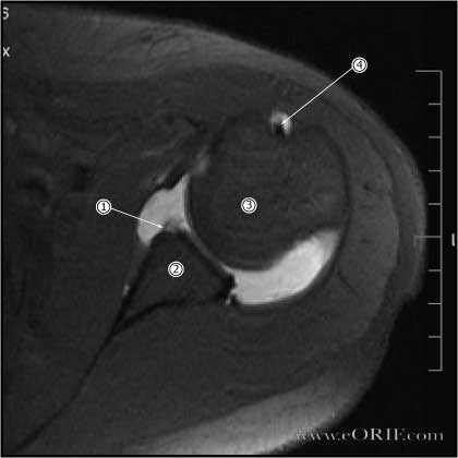
29yo RHD female with left shoulder anterioinferior instability. Initial sustained traumatic dislocation over 10 years earlier with several subsequent dislocations requiring sedation for relocation. At presentation dislocations occured with minimal abduction/ER movements. Gadolinium enhanced MRI shown.
|
 |
More inferior MRI image demonstrating bony Bankart lesion in the anterior inferior glenoid.
|
 |
30 y/o male 1 year after traumatic anterior-inferior shoulder dislocation with recurrent instability.
Arthroscopic view from beach chair position |
 |
Corresponding Hill-Sachs deformity in above patient.
Arthroscopic view from beach chair position |
 |
Example of Bankart repair using three 2.4mm Arthrex Biosuturetac anchors. |
