|
       
|
synonyms: Frozen Shoulder, adhesive capsulitis
Adhesive Capsulitis ICD-10
Adhesive Capsulitis ICD-9
- 726.0 (adhesive capsulitis of the shoulder)
- 719.51 (stiffness of joint not elsewhere classified involving shoulder region)
Adhesive Capsulitis Etiology / Epidemiology / Natural History
- Restricted active and passive shoulder motion in all planes. Pain and motion generally improve within 2 years.
- Generally late middle age (average 56). Common in IDDM patients. 16% bilateral involvement.
- Associated with diabetes, cervical spondylosis, hypothyroidism. Vascular endothelial growth factor (VEGF) may be involved in the pathogenesis in diabetic patients (Ryu JD, JSES 2006;15:679).
- Idiopathic possible causes: immunologic, inflammatory, biochemical, endocrine.
- Secondary adhesive capsulitis: known intrinsic, extrinsic, or systemic cause. Secondary causes: macrotrauma, microtrauma, or postsurgical intervention, combined with prolonged immobilization.
- 3 Stages:
Adhesive Capsulitis Anatomy
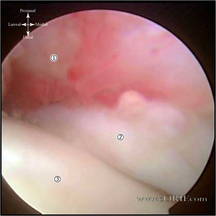
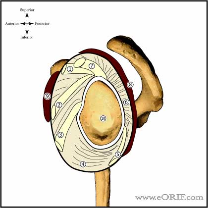
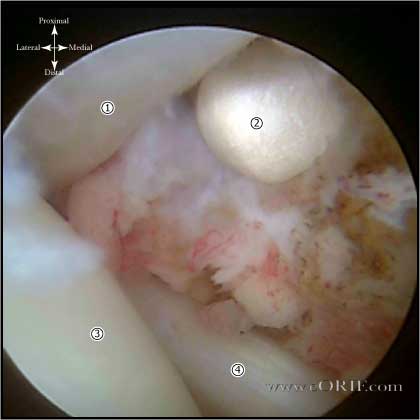
- Thickened, contracted joint capsule
- Decreased intra-articular volume
- Rotator interval and coracohumeral ligament contractures may be the most important lesions. (Ozaki J, JBJS 1989;71A:1511).
- "Essential lesion" is coracohumeral ligament contracture.
Adhesive Capsulitis Clinical Evaluation
- Keys= progressive pain and stiffness, spontaneous onset, decreased ROM in all planes, no tenderness, pain at extremes of motion.
- Both active and passive ROM limited due to soft-tissue contracture.
- Evaluate scapulothoracic motion.
- Consider evaluating AROM after subacromial lidocaine injection.
Adhesive Capsulitis Xray / Diagnositc Tests
- AP, scapular lateral and axillary views. Usually normal. May demonstrate osteopenia.
- Consider MRI to rule-out associated pathology. MRI demonstrates soft tissue thickening and increased signal intensity in the rotator interval obscuring the normal fat surrounding the coracohumeral ligament (coronal and sagittal oblique images). Also demonstrates abnormally thickened inferior glenohumeral ligament.
Adhesive Capsulitis Classification / Treatment
- Subacromial and glenohumeral injection (xylocaine &depomedrol) Q4wks x 3 with home stretching program. If fails to improve consider arthroscopic capsular release.
- Consider Medrol dose pack x 2. Take initial pack wait two weeks and then take second pack. Must be done with concomittant physical therapy.
- Idiopathic (primary)=usually responds to non-op treatment. Tends to resolve over 1-3 years. Glenohumeral corticosteriod injection can improve symptoms, especially if done early in the course of disease. Most will have some residual loss of motion over many years, but do not have functional limitations. Treatment options include supervised neglect, physical therapy, arthroscopic release, open release.
- Supervised neglect = explanation of the natural course of the disease, instructions not to exercise in excess of pain threshold, and instructed to do pendulum exercises and active exercises within this painless range and to resume all activities as tolerated = 89% near normal shoulder function at 24 months, 64% at 12 months. (Diercks, RL. JSES 13:499; 2004).
- Arthroscopic Capsular Release= Rotator interval release with circumferential capsular release for severe cases. RTC integrity remains intact allowing full active & passive ROM. Subacromial and subdeltoid contractures can be released(rare without prior surgery) (Warner JJ, JBJS 78A;1808:1996).
- Open capsular release allows subscap lengthening for pts with severe IR contracture. Allowing greater improvements in ER as compared to scope release. Subscapularis repair must be protected post-op (Omari A, JSES 2001;10:353).
- Collagenase injections: good results have been shown in early studies. Further evaluation is needed. (Badalemente M, AAOS Meeting 2006).
- Acquired (secondary)=postsurgical or posttraumatic= associated with prolonged immobilization= treatment is generally surgical release. Natural history is progressive arthritis especially with ER loss. Treatment = Arthroscopic Capsular Release
- Workers compensation patients have worse outcomes and frequently require MUA or capsular release. (Sean G, JBJS. 2000;82a:1398).
Adhesive Capsulitis Associated Injuries / Differential Diagnosis
Adhesive Capsulitis Complications
- Proximal humerus fracture
- Instability
- Stiffness / Arthrofibrosis
- Chondral Injury / Arthritis
- Infection
- RTC tear
- NVI (Axillary nerve palsy)
- Fluid Extravastion / Compartment Syndrome
- Complex Regional Pain Syndrome
- Synovial fistula
- Hemarthrosis / hematoma
- DVT / PE
- Recurrent pain and stiffness, Proximal humerus fracture, Instability, Stiffness / Arthrofibrosis, Chondral Injury / Arthritis, Infection, RTC tear, NVI (Axillary nerve palsy), Fluid Extravastion / Compartment Syndrome, Complex Regional Pain Syndrome, Synovial fistula, Hemarthrosis / hematoma, DVT / PE
Adhesive Capsulitis Follow-up Care
- Post-op: 48hours of intensive inpatient physical therapy with repeated interscalene regional analgesia or continuous infusion via an interscalene catheter.
Adhesive Capsulitis Review References
|


