|
|
synonyms: shoulder scope, SATS
Shoulder Scope CPT
Shoulder Scope Indications
Shoulder Scope Contraindications
Shoulder Scope Alternatives
- Open procedures
- Non-operative treatment
Shoulder Scope Pre-op Planning / Special Considerations
- Use 1:3000,0000 epinephrine irrigation fluid, electrocautery, hypotensive anesthesia(maintain SBP/subacromial BP difference of <50mm HG(Morrison DS, Arthroscopy 1995;11:557-60)
- Procedure may be done in beach chair or lateral postions.
- Beach Chair Position (BCP): intermittent SCD during surgery can increase cerebral oxygen saturation, cardiac output, stroke volume, ejection fraction, mean arterial pressure, and preload and decrease afterload. BCP Complications: hypotension, venous air embolism, cerebral and spinal ischemia, death. (Kwak HJ, Arthroscopy. 2010 Jun;26(6):729-33(, (Bickel A, Am J Surg. 2011 Jul;202(1):16-22.)
- Beach-chair position: cervical spine must be placed in a neutral position. Excessive pressure at the base of the skull in the region of the mastoid process can result in neurapraxia of the posterior auricular nerve. Excessive compression, flexion, extension, rotation, or lateral bending of the cervical spine can lead to brachial plexus injury, palsy of the hypoglossal nerve of superficial nerves of the neck. Excessive extension of the cervical spine can increase risk of stroke. The hips and knees should be flexed to decrease lumbosacral pressure, sciatic nerve tension and to prevent caudal migration of the patient.
- Shoulder arthoscopy beach chair drape: Halyard ref #89070.
Shoulder Scope Technique
- Pre-operative antibiotics, +/- interscalene block
- General endotracheal anesthesia
- Modified beach-chair position. All bony prominences well padded.
- Examination under anesthesia.
- Prep and drape in standard sterile fashion
- Bony landmarks marked out on the shoulder
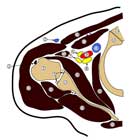
- Standard posterior portal just under posterior sulcus
- Anterior portal lateral to coracoid process. 18-gauge spinal needle utilized for localization with placement just under the biceps tendon in the rotator interval.
Examination Under Anesthesia
- Forward Elevation (R/L): 160 / 160
- External Rotation at side (R/L): 50 / 50
- External Rotation in 90 abduction (R/L): 85 / 85
- Internal Rotation in 90 adbuction (R/L): 20 / 20
- Load and shift
- Sulcus sign
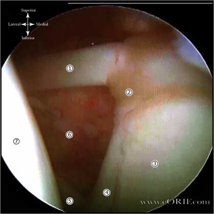
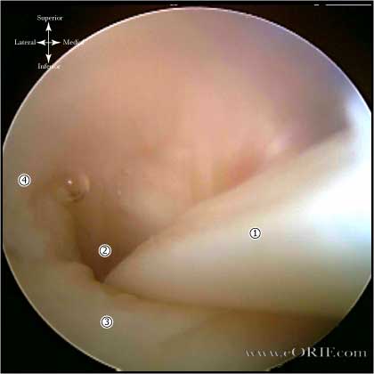
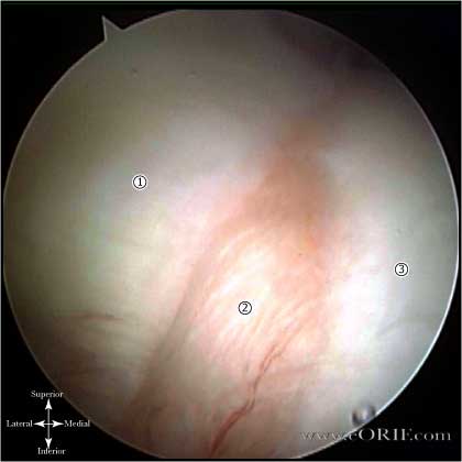
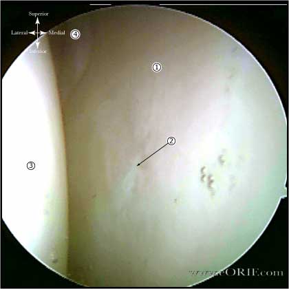
Viewing from Posterior Portal
- Biceps tendon, Biceps labral complex
- Biceps exit /
- Posterior superior labrum, posterior capsular recess
- Inferior axillary recess, anterior-inferior glenohumeral ligament
- Inferior labrum
- Glenoid articular surface
- Supraspinatus tendon: evaluate with arthrosope in posterior portal and arm elevated 50 degrees and internally rotated. This fascilites lesser tuberosity visualization. (Bennett WF Arthroscopy 2001;10:37). Only the proximal 25% of the subscapularis tendon can be visualized intra-articularly. (Wright JM, Arthroscopy 2001;17:677)
- Posterior rotator cuff / bare area
- Humeral articular surface
- superior, middle, inferior glenohumeral ligaments
- Subcapularis tendon
- Anterior inferior labrum, anterior inferior glenohumeral ligment
- Evaluate Peel-back sign = entire superior BLC will shift medially as the arm is ER while in 70-90 degrees of abduction. Generally found in posterior and combined anterior-posterior Type II SLAP lesions. (Burkhart SS, Arthroscopy 1998;14:637) (Burkhart SS, Arthroscopy 2003;19:404).
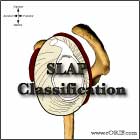
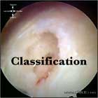
- RTC tear Classification, BLC classification, SLAP classification,
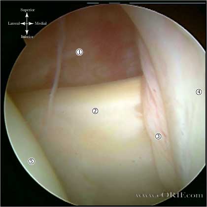
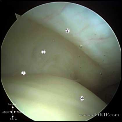
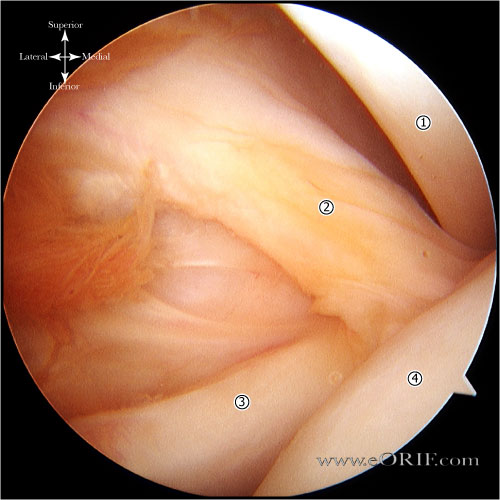
Viewing from Anterior Portal
- Posterior glenoid labrum
- Posterior capsule
- Posterior RTC (infraspinatus, terres
- Anterior inferior labrum
- Suscapularis tendon recess
- Middle glenohumeral ligament
- Subscapularis tendon, biceps tendon
Shoulder Scope Complications
- Infections
- Stiffness
- CRPS
- Nerve injury: Axillary nerve, Brachial plexus
- Fluid Extravasation:
- Chondrolysis: though to be related to heat from electo cautery or radiofrequency probes used during capsular release or capsular shrinkage.
- Hematoma
- Chondral Injury / arthritis
- DVT/PE
- Posterior auricular nerve neurapraxia: from exxcessive pressure at the base of the skull in the region of the mastoid process when using beach chair position.
- Infections, Stiffness, CRPS, Nerve injury: Axillary nerve, Brachial plexus, Fluid Extravasation, Chondrolysis, Hematoma, Chondral Injury / arthritis, DVT/PE
Shoulder Scope Follow-up care
- Post-op: sling as needed with pendulum ROM exercises.
- See Shoulder Arthroscopy Rehab Protocol.
- 1 week: Start PT focused on ROM and strengthening. AAROM, PROM. AROM, free weights start at 3 weeks. Avoid cross-body adduction for 6 weeks if DCR performed.
- 6 weeks: progressive sport specific activity.
- 3 months: Return to sport / full activities.
- IF in association with SAD, or RTC repair use those rehab protocols.
- Outcome measures: ASES score, pain scales.
Shoulder Scope Outcomes
Shoulder Scope Review References
- Moen, T, JAAOS 2014;22:410–419
- Burkhart SS, The Cowboy's Companion: A Trail Guide for the Arthroscopic Shoulder Surgeon, 2012
- Burkhart SS, Burkhart's View of the Shoulder: A Cowboy's Guide to Advanced Shoulder Arthroscopy, 2006
- AANA Advanced Arthroscopy: The Shoulder: Expert Consult: Online, Print and DVD, 1e, 2010
- Gartsman GM, Shoulder Arthroscopy, 2e, 2008
|


