|


|
synonyms: elbow fracture, olecranon Fracture
Olecranon Fracture ICD-10
Olecranon Fracture ICD-9
- 813.01(closed fracture of olecranon process of ulna)
- 813.11(open fracture of olecranon process of ulna)
Olecranon Fracture Etiology / Epidemiology / Natural History
- Common causes: MVA, falls directly onto posterior elbow
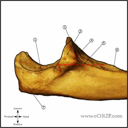
Olecranon Fracture Anatomy
Olecranon Fracture Clinical Evaluation
- Pain, swelling in elbow.
- Document NV exam.
- Document wrist evaluation.
Olecranon Fracture Xray / Diagnositc Tests
Olecranon Fracture Classification / Treatment
- Nondisplaced: sling immobilization followed by gentle ROM initiated within 10 days. Consider initially long arm casting for 4 weeks or ORIF depending of patient and fracture severity. Initially weekly xrays indicated to ensure fracture remains reduced.
- Displaced Young/high-demand: ORIF
- Displaced Elderly/low-demand: ORIF vs excision with triceps advancement.
- Olecranon Stress Fracture: generally associated with throwning sports, baseball. Treatment: rest and temporary splinting. Consider electrical stimulation. ORIF indicated after failure of nonoperative treatment. (Ahmad CS, Clin Sports Med 2004;23:665).
- Trans-olecranon fracture-dislocation: Treatment = ORIF. (Mortazavi SM, Injury. 2006 Mar;37:284, Ring D, JOT. 1997;11:545)
Olecranon Fracture Associated Injuries / Differential Diagnosis
Olecranon Fracture Complications
- Superficial wound infection
- Contracture
- Nonunion
- Hardware failure
- Malunion
- Painful hardware: occurs in 50%-80% of patients after olecranon fracture fixation. (tension band wiring frequently requires removal) (Karlsson MK, JSES 2002;11:377).
- Ulnar nerve palsy
Olecranon Fracture Follow-up Care
- Post-op: Place in posterior splint in 50-70º of flexion. Encourage finger ROM.
- 10-14 Days: Wound check, remove staples. Evaluate reduction on xrays. Place in long-arm cast or removable posterior splint depending on fracture type and strength of fixation.
- 6 Weeks: Wound check, evaluate xrays for callus formation. Place in removable posterior splint and begin elbow ROM exercises.
- 3 Months: Evaluate xrays for fracture union. Begin gentle sport specific exercises depending on fracture union. Consider bone stimulator if union not evident on xrays.
- 6 Months: Evaluate xrays. Return to sport.
- 1Yr: Evaluate xrays. Evaluate xrays using elbow outcome measures. May consider hardware removali f hardware is painful.
- (Bailey CS, JOT 2001;15:542).
- (Karlsson MK, CORR 2002;403:205).
Olecranon Fracture Review References
- Egol KA and Koval KJ in Fractures in Adults, 6th ed, 2006
- Rockwood and Green's Fractures in Adults: 2009
- Operative Elbow Surgery: Expert Consult: 2012

- AAOS/ASES Advanced Reconstruction Elbow, 2007

- Orthopaedic Knowledge Update: Shoulder and Elbow 3, 2008
- Veillette CJ, Steinmann SP. Olecranon fractures. Orthop Clin North Am. 2008 Apr;39(2):229-36
- Mortazavi SM, Asadollahi S, Tahririan MA. Functional outcome following treatment of transolecranon fracture-dislocation of the elbow. Injury. 2006 Mar;37(3):284-8.
- Ring D, Jupiter JB, Sanders RW, Mast J, Simpson NS. Transolecranon fracture-dislocation of the elbow. J Orthop Trauma. 1997 Nov;11(8):545-50.
|


