|

|
synonyms:radial head fracture ORIF
Radial Head ORIF CPT
Radial Head ORIF Indications
- Type II radial head fracture
- Elbow instability with associated radial head fracture
- Restriction of forearm rotation by radial head fragments
Radial Head ORIF Contraindications
- Type I radial head fracture
- Type III fracture in an elderly patient
Radial Head ORIF Alternatives
- Radial Head replacement
- Radial head Excision: may cause decreased grip strength, wrist pain, and progressive valgus instability
- Non-operative management
Radial Head ORIF Pre-op Planning / Special Considerations
- Equipement: Synthes mini fragment set, k-wires, +/- headless screws (Herbert, Accutrack) available
- Radial Head Plate Options
- Synthes Radial Head Plate
- Acumed Radial Head Plate
- Medartis Radial Head Plate
- Wright Medical EVOLVE® II Radial Head Plate
- Biomet Radial Head Plate
- Small Bone Innovations rHead Plating System
- Have prosthetic radial head available incase fixation proves inadequate.
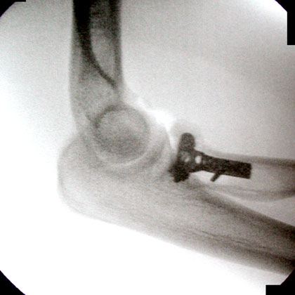
- C-arm
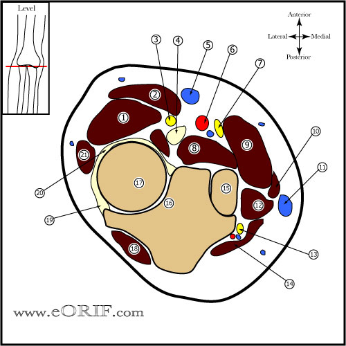
- Approach: standard Kocher approach (between ECU and anconeous) or direct full-thickness approach at the midline of the radiocapitellar joint. (Cohen MS, JAAOS 1998;6:15).
- Plate position for fixation of complex proximal radius fractures is with the forearm in neutral position, with the plate applied directly lateral. (Soyer Ad, JOT 1998;12:291)
- Safe zone: reference marks are made along radial head and neck so as to bisect the bone's anteroposterior distance. Three such marks are made with the forearm in neutral rotation, full supination, and full pronation. Next, the posterior limit of the zone is determined by bisecting the reference marks made with the forearm in neutral rotation and full pronation. The anterior limit is determined by going nearly two thirds of the distance from the neutral mark to that mark made in full supination. (Smith GR, JSES 1996;5:113).
- Safe zone for prominent fixation encompasses a 90° angle localized by palpation of the radial styloid and Lister's tubercle. (Caputo AE, J Hand Surg 1998;23:1082).
Radial Head ORIF Technique
- Pre-operative antibiotics, +/- regional block
- General endotracheal anesthesia.
- Supine position (with arm table, or arm can be brought across chest). All bony prominences well padded.
- Exam under anesthesia. Normal elbow ROM: Flexion/extension=0-135, Suppination=90, Pronation=90
- Tourniquet placed high on arm. Consider sterile tourniquet for complex injuries.
- Prep and drape in standard sterile fashion
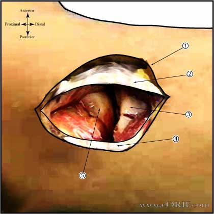
- Kocher lateral incision
- Interval between the anconeus and extensor carpi ulnaris opened. (Consider opening the white fascial layer which defines the extensor origin down to bone, going through the extensor origin, annular liagment and LCL complex as one unit.)
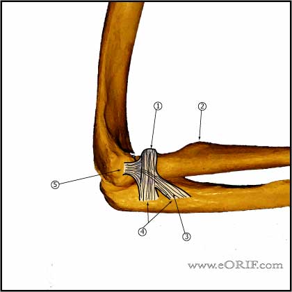
- ECU elevated exposing the lateral collateral ligament.
- Incise capsule anterior to the LCL complex.
- Evacuate hematoma / irrigate.
- Evaluated fracture pattern, remove any loose osteochondral fragments.
- Evaluate capitellum for osteochondral injury.
- Articular surface is reduced anatomically. Impacted areas are elevated. Consider provisional k-wire fixation.
- 2.0mm and 1.5mm screws or headless screws.
- If comminution extends down the neck 2.0mm or 2.4mm plate fixation is required. Plate position for fixation of complex proximal radius fractures is with the forearm in neutral position, with the plate applied directly lateral. (Soyer Ad, JOT 1998;12:291)
- Supinator is retracted/elevated from posterior to anterior with the forearm in pronation (protecting radial nerve) for plate placement. Exposure past the midline of the radial tuberosity risks PIN injury. (Tornetta P, 3rd, CORR 1997;215)
- Autologous bone graft can be obtained from olecranon process, distal radius or iliac crest.
- Evaluate for full elbow suppination / pronation and flexion and extension.
- If forearm is in neutral position, the safe zone is directly lateral. If forearm is fully supinated, the safe zone is posterior
- Irrigate.
- Close in layers. .
Radial Head ORIF Complications
- Stiffness, loss of ROM
- Failure of fixation
- Heterotopic ossification
- Instability (LCL injury)
- PIN palsy
- Radioulnar synostosis
- Nonunion
Radial Head ORIF Follow-up care
- Post-op: Splint with forearm in supination or neutral. Start early active range of motion as soon as possible. Consider Indomethacin 75mg QD/NSAIDs for patients with complex dislocations or Radial head replacement HO reduction.
- 7-10 Days: Evaluate incision, remove stitches, Begin early active range of motion as soon as possible. Consider hinged elbow brace for high energy injuries/instability. Start physical therapy. Avoid flexion in pronation. Patients with Essex-Loprestic injuries are held in full supination for 3-4 weeks.
- 6 Weeks: Consider static progressive nightime extension splinting if a flexion contracture is present 6 weeks after injury. 10° to 15° flexion contractures are not uncommon. If DRUJ treated by pinning remove pins at 6wks.
- 3 Months: Progress with ROM. May take 6-12 months to regain ROM. Begin sport specific therapy.
- 6 Months: May return to full activities provided patient is asymptomatic
- 1Yr: Assess outcomes, repeat xrays.
- Radial Head Rehab Protocol.
- See also Elbow Outcome Measures.
Radial Head ORIF Outcomes
Radial Head ORIF Review References
- Advanced Reconstruction-Elbow, AAOS 2007
- Hotchkiss RN, JAAOS 1997;5:1
|