|


|
synonyms: meniscal tear, meniscus tear
Meniscal Tear ICD-10
- S83.202 (A,D,S)- Bucket-handle tear of unspecified meniscus, current injury, unspecified knee
- S83.205 (A,D,S)- Other tear of unspecified meniscus, current injury, unspecified knee
- S83.209 (A,D,S)- Unspecified tear of unspecified meniscus, current injury, unspecified knee
- S83.219 (A,D,S)- Bucket-handle tear of medial meniscus, current injury, unspecified knee
- S83.229 (A,D,S)- Peripheral tear of medial meniscus, current injury, unspecified knee
- S83.239 (A,D,S)- Complex tear of medial meniscus, current injury, unspecified knee
- S83.249 (A,D,S)- Other tear of medial meniscus, current injury, unspecified knee
- S83.259 (A,D,S)- Bucket-handle tear of lateral meniscus, current injury, unspecified knee
- S83.269 (A,D,S)- Peripheral tear of lateral meniscus, current injury, unspecified knee
- S83.279 (A,D,S)- Complex tear of lateral meniscus, current injury, unspecified knee
- S83.289 (A,D,S)- Other tear of lateral meniscus, current injury, unspecified knee
- (A,D,S) A=initial encounter, D=subsequent encounter, S=sequela
Meniscal Tear ICD-9
- 717.0 old bucket handle tear of medial meniscus
- 717.1=derangement of anterior horn of medial meniscus
- 717.2=derangement of posterior horn of medial meniscus
- 717.3=other and unspecified derangement of medial meniscus
- 717.4=derangement of lateral meniscus
- 717.40=derangement of lateral meniscus unspecified
- 717.41=bucket handle tear of lateral meniscus
- 717.42=derangement of anterior horn of lateral meniscus
- 717.43=derangement of posterior horn of lateral meniscus
- 717.49=other and unspecified derangement of lateral meniscus
- 717.5=derangement of meniscus, not elsewhere classified (congenital discoid meniscus, etc)
- 836.0 (tear of medial cartilage or meniscus, current)
- 836.1 (tear of lateral cartilage or meniscus, current)
Meniscal Tear Etiology / Epidemiology / Natural History
- Most common knee injury
- Higher risk in ACL deficient knees
- Medial tears 3 times more common than lateral
- Risks of progressive arthritis are proportional to amount of meniscus removed.
- Traumatic tears generally caused by axial loading with rotation of a flexed knee.
- Male > female.
Meniscal Tear Anatomy
- Medial meniscus: C-shaped; anterior horn attaches ?; posterior horn attaches anterior to the insertion of the posterior cruciate ligament. (Johnson DL, Arthroscopy 1995;11:386) Average length = 45.7mm (McDermott ID, Knee Surg Sports Traumatol Arthrosc 2004;12:130). Secondary stabilizer to anterior tibial translation.
- Lateral Menisus: semicircular; anterior horn attaches adjacent to the ACL; posterior horn attaches behind the intracondylar eminence.(Johnson DL, Arthroscopy 1995;11:386) Average length = 35.7mm (McDermott ID, Knee Surg Sports Traumatol Arthrosc 2004;12:130).
- Discoid variant of the lateral meniscus is found in 3.5% to 5% of patients. (Vandermeer RD, Arthroscopy 1989;5:101)
- Average excursions of the menisci with knee flexion = 5.2 mm for the medial and 11 mm for the lateral meniscus. (Thompson WO, AJSM 1991;19:210)
- Provides shock absorption and attenuation of axial loads on the articular cartilage of the distal femur and proximal tibia. Secondary roles in knee stability, proprioception, joint lubrication and stress distribution.
- Medial meniscus transmits 50% of the load in the medial compartment. Lateral meniscus transmits 70% of the load in the lateral compartment. (Greis PE, JAAOS 2002;10:168). Degenerative changes occur earlier in pts undergoing parital lateral meniscectomy than partial medial meniscectomy. Menisci transmit more load in flexion (85%) than in extention (50%).
- Blood supply: the outer (peripheral) 20-30% of the medial meniscus and 10-25% of the lateral meniscus has blood supplied from the perimeniscal capillary plexus off the superior and inferior medial and lateral genicular arteries. (Arnoczky SP, AJSM 1982;10:90)
- Medial and lateral menisci are attached by the transverse (intermeniscal) ligament and posteriorly by the coronary ligaments
- 65% Water: 90% of collagen is type 1.
- see also Medvecky MJ, JAAOS 2005;13:121
Meniscal Tear Clinical Evaluation
- Pain and swelling in the knee. May have locking / catching / clicking sensation or giving way.
- May be associated with a twisting injury.
- Localized tenderness along the joint line.
- McMurray test: patient lying supine with the hip and knee flexed 90°. Apply axial compression while ER and IR the leg. Reproduction of pain +/- clicking indicates meniscal tear.
- Apley Compression Test
- Ege's test
- Note.
Meniscal Tear Xray / Diagnostic Tests
- Knee xrays are generally normal but are indicated to evaluate for arthritis and rule out concomitant disorders.
- MRI: tears and intrasubstance degeneration appear as marked increased signal on T1 and intermediate weighted images that is less prominent on T2 images. Grade 1 = poorly defined globular zone of increased signal. Grade 2 = a linear zone of hyperintensity not communicating with free meniscal surface. Grade 3 = a linear band of hyperintensity commonicating with a freee meniscal surface. (Stoller DW, Radiology 1988;166:580). Extrusion of the meniscus, defined as >3mm measured from the outer magrin of the meniscus to the outer articular margin of the medial tibial plateau on mid coronal MRI scans, is associated with posterior horn avulsion/meniscal root tear (Marzo JM, JAAOS 2009;17:276).
- MRI Insurance Indications: acute injury with mechanical symptoms (locking, popping). Chronic mechanical symptoms that has failed 4 weeks of PT, or has mechanical symptoms in which an intraarticular loose body in the differential diagnosis.
- “Double PCL” sign indicates a displaced bucket-handle tear of the medial meniscus into the intercondylar notch. Bucket-handle tear of the lateral meniscus generally appears in the anterior aspect of the lateral compartment.
Meniscal Tear Classification / Treatment
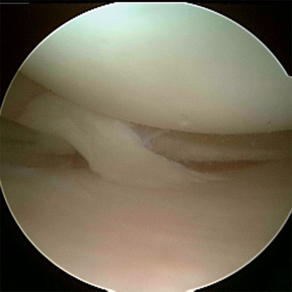
- Classification: longitudinal, bucket handle, flap, radial, horizontal cleavage, Complex/degenerative. Meniscal root avulsion.
- Vascularity: red-red(within 3mm of meniscosynovial juntion) vs red-white(3mm-5mm from meniscosynovial junction) vs white-white(>5mm from meniscosynovial junction)
- Treatment: depends on tear pattern and location, see specific tear type.
- Non-op: activity modification or elimination of painful activities, ice, compression anti-inflammatory or analgesic medications. Weight loss in obese patients. Consider soft shock-absorbing shoe wear, medial-post orthotics, or valgus unloader brace. (Marzo JM, JAAOS 2009;17:276)
- Partial Meniscectomy: 94.8% Good/excellent results for isolated meniscal tear, 62% good/excellent results for patients with additional cartilage damage. (Schimmer RC, Arthroscopy 1998;14:136).
- Mensical Repair
- Meniscal Transplantation
- Discoid Meniscus
Meniscal Tear Associated Injuries / Differential Diagnosis
- Knee Arthritis
- LCL, Posterolateral cornercombined injury
- MCL: need for concurrent repair is dependent on amount of valgus laxity. 2-3mm increased laxity is probably not detrimental. Absolute indication for MCL repair has not been established.
- Chondral injury.
- PCL tear
- ACL tear: Shelbourne’s showed a decrease in post operative loss of extension in patients treated with meniscal repair followed by delayed ACL reconstruction compared to those treated with simultaneous repairs for patients with locked bucket-handle meniscal tears and chronic ACL deficiency. (Shelbourne KD Am J Sports Med 1993;21:779). Isolated stable tears of the posterior horn of the lateral meniscus posterior to the popliteus can be left untreated during ACL reconstruction.
- Patellar Dislocation
- Popliteus avulsion
- Knee Dislocation
- Tumor (Muscolo DL, JBJS 2003;85A:1209).
- Discoid Meniscus
Meniscal Repair Complications
- Overall complication rate = 1.8% (Small NC, Arthroscopy 1988;3:215)
- DVT: 9.9%, proximal DVT rate = 2.1% (Ilahi OA, Arthroscopy, 2005;21:727)
- Stiffness / Arthrofibrosis
- Chondral Injury / Arthritis
- Infection
- NVI (medial = saphenous neuralgia, lateral = peronela nerve palsy)
- Fluid Extravastion / Compartment Syndrome
- Complex Regional Pain Syndrome: rare
- Hemarthrosis
- Synovial fistula
- DVT, Stiffness / Arthrofibrosis, Chondral Injury / Arthritis, Infection, NVI (medial = saphenous neuralgia, lateral = peronela nerve palsy), Fluid Extravastion / Compartment Syndrome, Complex Regional Pain Syndrome, Hemarthrosis, Synovial fistula
Meniscal Repair Follow-up
- 80-90% successful.
- Hinged-knee brace with 0 to 90° of motion, 25% weight bearing with crutches for 4 wks.
- Full weight bearing and flexion beyond 90° usually is allowed after 6 to 12 weeks postoperatively.
- Squating is prohibited for 4 months.
- Patients may return to sedentary work at 1 week, strenuous work at 3 to 4 months, low-impact exercises at 8 weeks, and running after 4 to 5 months. Light or moderate sports are allowed at 6 to 9 months.
Meniscal Tear Review References
|


