 |
synonyms: MCL tear, MCL rupture, MCL sprain, medial collateral ligament tear, tibial collateral ligament
MCL ICD-10
MCL ICD-9
- 844.1 (sprain and strains of knee and leg; medial collateral ligament of knee)
- 717.82 (old disruption of medial collateral ligament)
MCL Etiology / Epidemiology / Natural History
- Typically results from a valgus contact injury.
- Prophylaxis: knee bracing decreases the risk of MCL injury, but does not affect ACL or LCL injuries (Najibi S, AJSM 2005;33:602).
MCL Anatomy
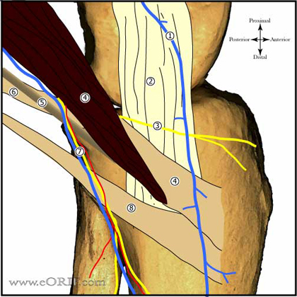
- Composed of superficial and deep fibers. (Satterwhite Y, Op Tech Sports Med 1996;4:134) (Warren RF, JBJS 1979;61A:56). MCL complex = superficial MCL, deep MCL and posterior oblique ligament.
- Origin: Superficial MCL=medial femoral epicondyle
- Insertion: Superficial MCL=periorsteum of proximal tibia, deep to the pes anserinus.
- Function: Primary restraint to valgus instability; secondary stabilizer to anterior translation; also resists external rotation.
- Blood Supply:
- Deep MCL is a capsular thickening.
- More commonly injury at is femoral origin than tibial insertion.
MCL Clinical Evaluation
- Medial knee tenderness, often medial epicondyle. Local swelling is common. Not usually associated with large effusion unless concomitant ligament injury is present.
- Valgus laxity with the knee in 30° of flexion as compared to normal side indicates isolated MCL tear. Laxity in full extension is indicative of a more severe injury involving multiple ligaments and potentially both cruciates.
MCL Xray / Diagnositc Tests
- A/P and lateral knee xrays indicated. Generally normal in isolated MCL injuries.
- Stress xrays are indicated in pediatric patients to rule out distal femoral physeal fracture.
- Pellegrini-Stieda lesion: calcification adjacent to the medial femoral epicondyle seen in chronic MCL injuries.
- MRI: Grade 1 = edema (increased T2 signal) superficial to intact ligament. Grade 2 = edema within the substance of the ligament with some intact fibers. Grade 3 = complete ligament disruption (high signal traversing ligament on T2 images. (Schweitzer ME, Radiology, 1995;22:411). MCL tears with displacement intraarticularly on MRI require repair. Injuries over the entire length of the MCL are associated with residual valgus laxity with brace treatment (Nakamura H, AJMS 2003;31:261). Often demonstrate bone marrow edema in the lateral femoral condyle and Lateral tibial plateau.
MCL Classification / Treatment
- Acute Isolated MCL injury:
Grade I injury (0 to 5 mm of medial opening)
Grade II injury (6 to 10 mm of medial opening)
Grade III injury (11 to 15 mm of medial opening).
Treatment: WBAT, Hinged Knee brace with early motion and functional rehabilitation. Typically brace opened from 15° to 60° for first 4weeks. Then open 0°-120°for 4 weeks for a total of 8 weeks of bracing. (Indelicato PA, JBJS 1983;65A:323). Consider ice, compression, NSAIDs
- Chronic MCL injury which has failed non-operative treatment. Treatment: MCL reconstruction. Concomitant osteotomy indicated if valgus deformity is present on limb alignment xrays.
- ACL/MCL injury = WBAT, Hinged knee brace with early motion and functional rehabilitation. Typically brace opened from 15° to 60° for first 4weeks. Then open 0°-120°for 4 weeks for a total of 8 weeks of bracing. ACL reconstruction performed after full ROM restored. (Petersen W, Arch Orthop Trauma Surg 1999;119:258), (Halinen J, AJSM 2006;34:1134). Concomitant MCL reconstruction indicated if valgus laxity persists after conservative treatment.
- PCL / MCL injury = if posterior subluxation is present acute MCL reconstruction and PCL reconstructionis indicated. If joint is reduced and stable MCL is treated with bracing and PCL is reconstructed when valgus stability and full ROM are restored.
- Acute Multiple ligament knee injury / Knee Dislocation
Treatment: MCL reconstruction, and concomitant ligament repair/reconstruction.
- MCL Tear Patient Information
MCL Associated Injuries / Differential Diagnosis
MCL Complications
- Instability
- Stiffness:
- Painful hardware:
- Infection:
- Arthrofibrosis: rare
- NVI (saphenous neuralgia): rare
- Complex Regional Pain Syndrome: rare
- Hemarthrosis
MCL Follow-up Care
- Return to play is based on pain and residual instability. Average time to return to play: Grade I=2 weeks; Grade II=3 weeks; Grade III=6 to 8 weeks.
MCL Review References
|

