|

|
synonyms: coronoid fracture, elbow fracture-dislocation
Coronoid Fracture ICD-10
- S52.043A Displaced fracture coronoid process unspecified ulna, initial closed
- S02.630A Fracture coronoid process mandible, unspecified side, initial closed
- S52.044A Nondisplaced fracture coronoid process right ulna, initial closed
- S02.631A Fracture coronoid process right mandible, initial closed
- S52.045A Nondisplaced fracture coronoid process left ulna, initial closed
- S02.632A Fracture coronoid process left mandible, initial closed
- S52.046A Nondisplaced fracture coronoid process unspecified ulna, initial closed
- S52.041A Displaced fracture coronoid process right ulna, initial closed
- S52.042A Displaced fracture coronoid process left ulna, initial closed
- SEE ALL CORONOID PROCESS FRACTURE ICD-10
Coronoid Fracture ICD-9
- 813.02(closed fracture of coronoid process of ulna)
- 813.12(open fracture of coronoid process of ulna)
Coronoid Fracture Etiology / Epidemiology / Natural History
- Occur in 2-10% of elbow dislocations
Coronoid Fracture Anatomy
Coronoid Fracture Clinical Evaluation
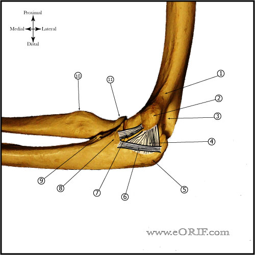
- Evaluate for tenderness or bruising at the radial and ulnar collateral ligament origins.
- Evaluate for elbow stability.
- Document NV exam.
- Document wrist evaluation.
Coronoid Fracture Xray / Diagnositc Tests
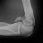
- A/P, lateral and oblique elbow films indicated. Coronoid fractures are often obscurbed by associated radial head fracture. "River delta sign"=narrowing of the joint space from lateral to medial=indicates coronoid fx or ligamentous instability.
- Consider stress xrays to determine ligamentous stability. If any concern for instability is present stress radiographs are indicated. Stress xray comparisons to uninjured side are always helpful.
- CT is best for determining fracture location and comminution and should be considered if the diagnosis is questionable, especially if associated with radial head fracture.
Coronoid Fracture Classification / Treatment
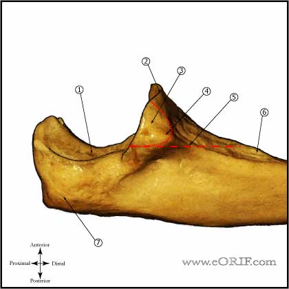
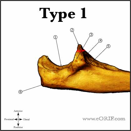
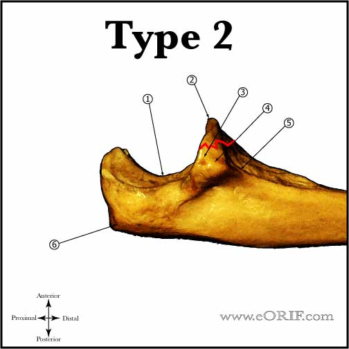
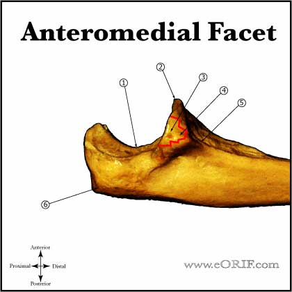
- Reagan and Morrey (JBJS 71A:1348, 1989) classification based on review of 35 patients. Anteromedial Facet fractures later described by O'Driscoll.
- Type I, stable (avulsion of the tip of the coronoid process): usually related to posterolateral rotatory elbow subluxation. Treatment = early protected ROM.
- Type I, unstable or associated radial head fracture. Treatment = ORIF; if fragment is large enough for fixation with a screw or k-wire it should be fixed via the lateral exposure to the radial head. Any collateral ligament injury must be repaired as well.
- Type II, stable( <50% of coronoid): Treatment = early protected ROM.
- Type II, unstable or associated radial head fracture. Treatment = ORIF; if fragment is large enough for fixation with a screw or k-wire it should be fixed. Any collateral ligament injury must be repaired as well.
- Type III (basal coronoid fracture): Treatment = ORIF usually via a posteromedial aproach. Often associated with olecranon fracture/dislocations. Associated injuries should be anatomically repaired as well.
- Anteromedial Facet Fracture, stable: Occur with varus posteromedial rotation during axial loading. Associated with LCL rupture and are usually unstable. Treatment = ensure joint is stable with stress radiographs, consider EUA. Early protected ROM if joint is confirmed to be stable.
- Anteromedial facet fracture, unstable: Treatment = ORIF with concomitant LCL/radial head repair usually via a utilitarian posterior exposure with posteromedial coronoid exposure. (Doornberg JN, JBJS 2006;88A:2216).
- Always consider hinged external fixation for severe injuries where joint stability is a concern post-operatively.
Coronoid Fracture ORIF Technique
- Note: Coronoid fractures can be fixed via a posteromedial exposure, or from a posterolateral exposure during radial head repair. A utilitarian posterior incision is general used.
- Pre-operative antibiotics
- Supine with arm board, tourniquet high on arm, C-arm available
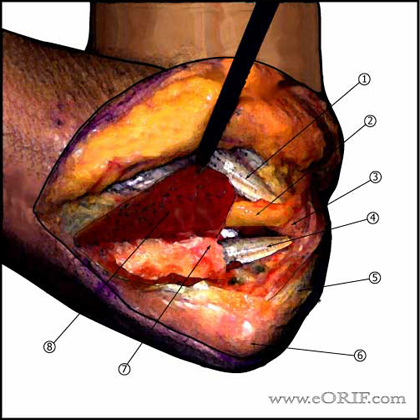
- Posterior incision from 6cm proximal to olecranon to 6cm distal to olecranon. Curved around medial border of olecranon to avoid painful scar.
- Medial and lateral skin flaps are raised depending on associated pathology.
- Ulnar nerve identified and transposed anterior to the medial epicondyle.
- Flexor carpi ulnaris incised longitudinally, leaving a fascial cuff for later repair. FCU is subperiosteally elevated exposing anterior band of medial collateral ligament and the coronoid.
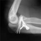
- Fracture is repair using AO techniques with k-wires, screws and buttress plate as needed.
- Irrigate.
- FCU repaired.
- Ulnar nerve secured anterior to the medial epicondyle with a fascial sling.
- Close in layers.
- Consider the use of a unilateral hinged external fixator placed on the lateral side to counteract varus gravitational stresses.
Coronoid Fracture Associated Injuries / Differential Diagnosis
Coronoid Fracture Complications
- Instability
- Stiffness, most will experience some loss of extention.
- Arthritis
- Infection
- Heterotopic ossification
Coronoid Fracture Follow-up Care
- Bulkly dressing with posterior splint post-operatively
- 7-10 day post-operative: Splint removed, ROM in a hinged elbow brace is started with ROM determined by security of fixation achieved at surgery.
- See also Elbow Outcome Measures.
Coronoid Fracture Review References
|


