synonyms: core decompression,
Core Decompression Humeral Head CPT
Core Decompression Humeral Head Indications
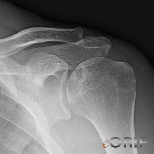
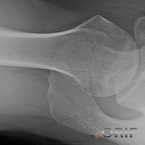
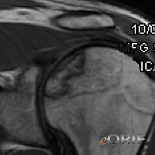
- Humeral head osteonecrosis (avascular necrosis) stage I and II
Core Decompression Humeral Head Contraindications
- Humeral head osteonecrosis Stage III, IV, V
Core Decompression Humeral Head Alternatives
- Conservative care (PT, activity modification, NSAIDs)
- Humeral head resurfacing
- Humeral hemiarthroplasty
- Total Shoulder replacement
Core Decompression Humeral Head Pre-op Planning
- Consider Wright Medical core decompression kit and graft
Core Decompression Humeral Head Technique
- 2cm stab incision for access. Under fluoroscopic guidance (both AP & Lat. views), introduce the 3.2mm fluted guide-wire into central aspect of lesion.
- Place tissue protector over the guide-wire down to bone.
- Using 9mm cannulated drill bit, decompress the femoral head
- Place working cannula with obturator through the tissue protector and into the core.
- Remove the tissue protector and obturator.
- Place X-REAM™ Percutaneous Expandable Reamer. Cofirm placement with fluoro (AP & Lat. views). Turn the blade control knob ¼ turn (or less) clockwise and rotate the entire instrument at least one full revolution. Repeat until desired expansion is achieved. Frequently confirm blade position under fluoro (BOTH A/P and LAT views).
- Removed X-Ream device. Irrigate and suction.
- Backfill the core with bone graft or Injectable Graft.
- Close in standard fashion.
Core Decompression Humeral Head Complications
Core Decompression Humeral Head Follow-up care
- Post-op: sling, pendelum ROM exercises
- 7-10 Days: Follow up for wound check and start PT
- 6 Weeks: Xrays, assess ROM, gradually resume normal activities.
- 3 Months: Xrays, progress with activities as tolderated.
- 6 Months: Xrays, evaluate function and ROM.
- 1Yr: Xrays.
Core Decompression Humeral Head Outcomes
Core Decompression Humeral Head Review References


