|


|
synonyms: colar bone fracture
Clavicle Fx ICD-9
- 810.01 (closed fracture of sternal end clavicle)
- 810.02 (closed fracture of shaft clavicle)
- 810.03 (closed fracture of acromial end clavicle)
- 810.11 (open fracture of sternal end clavicle)
- 810.12 (open fracture of shaft clavicle)
- 810.13 (open fracture of acromial end clavicle)
Clavicle Fx Etiology / Epidemiology / Natural History
Clavicle Fx Anatomy
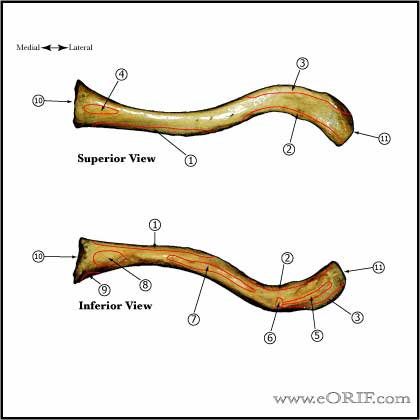
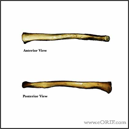
- S shaped: anterolateral surface is concave, convex on anteromedial surface.
- First bone to ossify, 5th gestational week
- Only long bone to ossify be intramembranous ossification
- Medial physis=80% of longitudinal growth
- Both acromial and sternal physes can remain open into 3rd decade of life
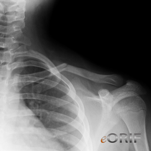
- Middle 1/3 fractures typically heal with the lateral shaft fragment displaced posterior to the medial shaft fragment / anterior-posterior angulation. (Edleson JG, JSES 2003;12:173).
- Diameter of medullary canal is approximately 7mm at its narrowest point. (Andermahr J, Clin Anat 2006;20:48).
- See also Shoulder Anatomy.
Clavicle Fx Clinical Evaluation
- Pain over clavicle, crepitus and motion at fracture site. Often obvious deformity.
- Evaluate skin over fracture site for tenting, inpending skin compromise
- Ptosis of affected shoulder.
- NV exam of upper extremity indicated; rule out axillary artery/vein injury.
- Auscultate chest to rule out associated pneumothorax.
- Medial clavicle fractures may have dysphagia and shortness of breath.
Clavicle Fx Xray / Diagnositc Tests
- A/P view of clavicle and 30°-45º cephalic tilt view of clavicle.
- Apical oblique (Grashe with 20 cephalad)
- Abduction Lordotic (after ORIF)
- Serendipity view (helps with A/P displacement)
- Axillary
- Zanca 15 degree apical oblique (AC joint)
- Consider chest xray (including both clavicles) if there is significant deformity (shortening) or if concerned for pneumothorax or rib fx's.
- CT with 3-D reconstruction is helpful for segmental fractures or for medial clavicle fractures which may be missed on Chest xrays.
Clavicle Fx Classification / Treatment
- Edinburg Classification (Robinson CM, JBJS 1998;80Br:476)
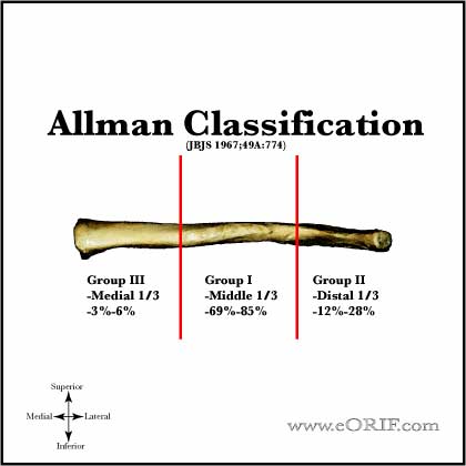
- Allman Classification (Allman FL, JBJS 49A;774:1967).
- Group I - middle 1/3=69%-85%
-69%-85% of clavicle fractures.
-0.13%-13% non-op nonunion rate overall
-Non-displaced: activity modifications, sling. No difference between sling and figure-8 (Stanly, Injury 19;162:1988), (Andersen K, Acta Orthop Scan 1987;58:71). Figure-8 has been associated with increased axillary pressure sores, compression of the neurovascular bundle, nonunion and patient discomfort. Closed reduction not helpful (Hill, JBJS 1997;79B:537).
-Displaced: generally treated conservatively with a sling. Large potential for remodeling.
Clavicle Fx Associated Injuries / Differential Diagnosis
Clavicle Fx Complications
- Nonunion
- Malunion: principle deformity is anterior angulation.
- Infection
- Neurovascular Injury
- Hardware failure (Brouwer KM, JSES 2008 IP)
- Sequelae of clavicle fractures: shoulder weakness, fatigue, paresthesias, cosmetic deformity, pain at rest or with activity, asymmetry.
Clavicle Fx Follow-up Care
- Post-op: sling for comfort, no overhead motion. Immediate pendelum ROM exercises.
- 10-14 Days: Wound check, sutures removed. Start PT for gentle ROM exercises. No resistive exercises/activities. Sling as needed for comfort.
- 6 weeks: Xrays, if union is evident begin strengthening and resistive exercises. No contant athletics.
- 3 months: Repeat xrays. If no signs of union, consider bone stimulator, see Nonunion.
- Return to sport: there shoulde be both clinical and radiographic evidence of union. Fracture site should be nontender, full shoulder ROM achieved and strength comparable to contralateral side. Generally avoid contact sports and heaving lifting for 4-6 months.
- Nonoperative outcomes: (Robinson CM, JBJS 2004;86A:778).
- Mean time to union = 28.4 weeks for non-operative treatment and 16.4 weeks for operative treatment for displaced clavicle fractures. (McKee MD, JBJS 2007;89A:1).
- Shoulder Outcome measures.
Clavicle Fx Review References
|