| See also Tibial Plateau Fracture ICD-10 Classification S82.109A |
|
 |
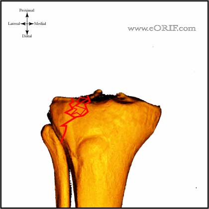
Type I
- “pure cleavage”, split lateral plateau; generally younger pts, high risk of ligament injury (MCL), or trapped meniscus
- Nondisplaced (<2mm): Non-weighted bearing in knee immobilizer or cylinder cast for 7/10 days. Repeat films at follow-up, if stable switch to hinged- knee brace with toe touch weight-bearing x 8-12wks and isometric quad exercises with progressive passive/active assist ROM. (Segal D, CORR 1993;294:232) (DeCoster TA, CORR 1988;231:196)
- Displaced (>2mm, >10 degree angulation): ORIF vs CRPP, generally ligamentotaxis reduction and fixation with 6.5 or 7.0mm cancellous lag screws. Treatment is depended on the condition of the soft tissues. Consider spanning external fixation with delayed ORIF when soft-tissues permit for severe soft-tissue injury.
|
 |
Type II
- “cleavage with depression”, split depressed lateral plateau fx
- Nondisplaced (<2mm):
- Displaced (>2mm, >10 degree angulation): ORIF. Generally ligamentotaxis reduction with depressed area elevated with bone tamps and packed with graft through fracture site or cortical window and fixation with peri-articular plate with multiple 3.5mm subchondral cortical "raft" screws proximally (Mills WJ, Orthop Clin North AAM 2002;33:177). Treatment is depended on the condition of the soft tissues. Consider spanning external fixation with delayed ORIF when soft-tissue permit for severe soft-tissue injury
|
 |
Type III
- “pure central depression” of lateral plateau, lateral cortex intact; older pt, low risk of ligament injury
- Nondisplaced (<2mm): Non-weighted bearing in knee immobilizer or cylinder cast for 7/10 days. Repeat films at follow-up, if stable switch to hinged- knee brace with toe touch weight-bearing x 8-12wks and isometric quad exercises with progressive passive/active assist ROM. (DeCoster TA, CORR 1988;231:196)
- Displaced (>2mm, >10 degree angulation): ORIF. Depressed area elevated with bone tamps and packed with graft through fracture site or cortical window and fixation with 6.5 or 7.0mm cancellous lag screws. Treatment is depended on the condition of the soft tissues. Consider spanning external fixation with delayed ORIF when soft-tissues permit for severe soft-tissue injury.
|
 |
Type IV
- medial plateau fx; high-energy, associated with traction injury to peroneal nerve
- Nondisplaced (<2mm):Non-weighted bearing in knee immobilizer or cylinder cast for 7/10 days. Repeat films at follow-up, if stable switch to hinged- knee brace with toe touch weight-bearing x 8-12wks and isometric quad exercises with early ROM. (DeCoster TA, CORR 1988;231:196)
- Displaced (>2mm, >10 degree angulation): ORIF. Generally ligamentotaxis reduction and fixation with peri-articular plate with multiple 3.5mm subchondral cortical screws proximally. Incisions determined by pre-operative CT. Plae should be placed with base over the apex of the fracture. Treatment is depended on the condition of the soft tissues. Consider spanning external fixation with delayed ORIF when soft-tissue permit for severe soft-tissue injury.
|
 |
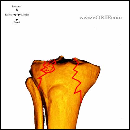
Type V
- Bicondylar.
- Treatment = ORIF. Generally ligamentotaxis reduction fixation with lateral peri-articular locking plate with multiple 3.5mm subchondral cortical screws proximally. Consider separate medial incision and plating. Treatment is depended on the condition of the soft tissues. Consider spanning external fixation with delayed ORIF when soft-tissue permit for severe soft-tissue injury.
|
 |
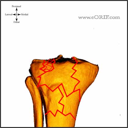
Type VI
- Plateau fractures with dissociation of metaphysis and diaphysis.
- Treatment ORIF. Generally ligamentotaxis reduction fixation with lateral peri-articular locking plate with multiple 3.5mm subchondral cortical screws proximally. Consider separate medial incision and plating. Treatment is depended on the condition of the soft tissues. Consider spanning external fixation with delayed ORIF when soft-tissue permit for severe soft-tissue injury.
|


