Syndesmosis Fixation 27829


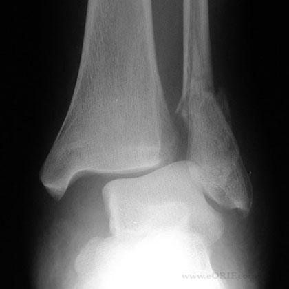
Ankle syndesmosis injury xray 

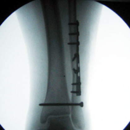
Ankle syndesmosis fixation xray

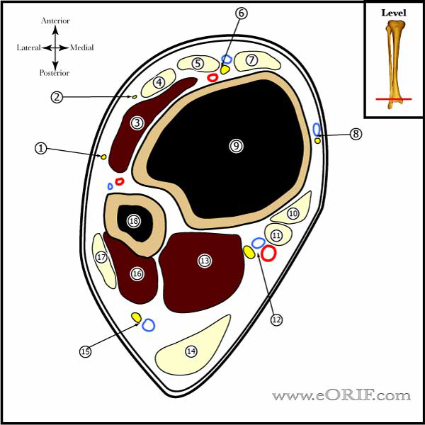
Ankle Cross section image

Lateral malleolus fracture syndesmosis injury xray

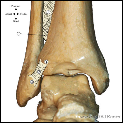
Ankle syndesmosis image

Lateral Malleolus fracture ORIF xray operative fixation


synonyms: syndesmosis fixation

Syndesmosis Fixation CPT

Syndesmosis Fixation Indications

  • Syndesmosis instability after fixation of associated fractures.
  • Syndesmotic injury with diastasis or instability.

Syndesmosis Fixation Contraindications

  • Syndesmotic injury without diastasis or instability.

Syndesmosis Fixation Alternatives

  • Nonoperative treatment.

Syndesmosis Fixation Planning / Special Considerations

  • Dynamic syndesmotic fixation has improved clincal outcome and decreased complications compared to static screw fixation. (Grassi A, AJSM 2020;48(4):1000–1013)
  • Dynamic syndesmotic fixation options: Arthrex Tightrope, Wright Medical Gravity Synchfix or Biomet toggleloc devices.  
  • When using dynamic fixation.  Consider making 1-cm medial approach prior to drilling or passing the suture button due to high risk of injury to the saphenous vein and/or nerve.  Consider double fixation with fixation at divergent angles for additional fibular rotational control.  (Berlet GC, Orthopedicstoday, 2018, Feb)Fixation can be provided with two tricortical 3.5-mm screws or a 4.5-mm polylactide bioabsorbable screw, a 4.5mm stainless steel screw, or dynamic flexible fixation. Dynamic syndesmotic fixation may have faster recovery without need for removal.
  • Consider the degree of instability and the size of the fibula when selecting screw size and determining the number of cortices engaged.
  • For high fibula fractures consider ORIF of the fibula to aid in determining proper rotation and length during syndesmotic fixation.

Syndesmosis Fixation Technique

  • Sign operative site.
  • Pre-operative antibiotics, +/- regional block.
  • General endotracheal anesthesia
  • position. All bony prominences well padded.
  • Examination under anesthesia.
  • Prep and drape in standard sterile fashion.
  • Position the ankle in neutral. (Ankle position is controversial).
  • Reduce syndesmosis with a periarticular reduction forceps, if needed, do not overtighten.
  • Centered drill on the fibula to avoid fracture.
  • Consider Arthrex Tightrope technique, Wright Medical Gravity Synchfix or Biomet toggleloc devices.  
  • Drill tricortical or quadcortical hole using standard AO technique 2-4cm proximal and parallel to the ankle joint in the coronal plane.
  • In the transverse plane, aim 25° to 30° obliquely from posterolateral to anteromedial because the fibula is located posterior to the tibia.
  • Place 4.5mm stainless steel, fully threaded screw using AO technique securing the syndesmosis. Do not insert screw in lag fashion.
  • Irrigate.
  • Close in layers.

Syndesmosis Fixation Complications

  • Instability
  • Broken hardware
  • Painful hardware
  • Pain
  • Syndesmosis ossification
  • Arthritis
  • Infection
  • Wound breakdown
  • Instability, Broken hardware, Painful hardware, Pain, Arthritis, Infection

Syndesmosis Fixation Follow-up care

  • Post-op: place in NWB Bulky-Jones dressing with posterior mold and side splints.
  • 7-10 Days: Wound check. Place in NWB cast.
  • 6 Weeks: Cast removed, check xrays. Place in Cam-walker with slow progression to full weight bearing. Discontinue cam-walker if pain free after 4 weeks.
  • 3 Months: schedule for syndesmotic screw removal to be done 12weeks after placement.
  • 6 Months: Resume activities.

Syndesmosis Fixation Outcomes

Syndesmosis Fixation Review References

  • Zalavras C, JAAOS, 2007;15:330
  • Grassi A, AJSM 2020;48(4):1000–1013