|


|
synonyms: forearm fracture ORIF, Radius and Ulnar Shaft Fracture ORIF, forearm fracture ORIF
Forearm Fracture ORIF CPT
Forearm Fracture ORIF Indications
- Displaced forearm fracture in an adult
Forearm Fracture ORIF Contraindications
- Active infection
- Gross contamination
- Severe soft tissue injury
Forearm Fracture ORIF Alternatives
- External Fixation
- Intramedullary nails
- Casting
Forearm Fracture ORIF Pre-op Planning
- Consider Acumed Precountoured Radius and Ulna Shaft Plates, IM nails
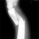
- Must ensure the radial bow is restored. Malrotation results in loss of pronation / supination.
- Primary bone grafting for comminuted fractures has not been shown to improve union rates. (Ring D, J Trauma 2005;59:436).
- Template case base on pre-operative xrays to ensure appropriate plate size and placement.
- AO Classification with approaches
Forearm Fracture ORIF Technique
- Sign operative site.
- Pre-operative antibiotics, +/- regional block.
- Supine position. Hand table; tourniquet high on arm; all bony prominences well padded.
- General endotracheal anesthesia
- Prep and drape in standard sterile fashion. Arm exsagninated with Esmarch, tourniquet inflated
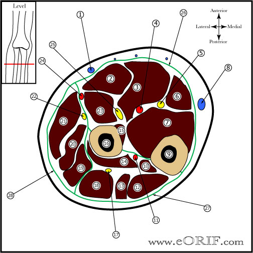
- Radius= Henry volar approach. Distally develop plain between brachioradialis and FCR.
- Ensure superficial sensory branch of radial nerve is preseved distally and PIN is preserved proximally.
- Minimize periosteal stripping and soft tissue dissection as much as possible
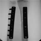
- Expose fracture, reduce and plate using standard AO technique. Generally 8-cortices above and below fracture. For purely transverse fractures 6-cortices (6 hole plate) is effective.
- Consider dorsal Thompson approach for fx of the proximal 1/3 of the radius. Plane between EDC and ECRB. Elevate suppinator with forearm in supination to protect PIN
- Ulnar fixation via straight dorsal approach along subcutaneous border of ulna. Place plate dorsal surface of ulna beneath the ECU
Close in layers.
Forearm Fracture ORIF Complications
- Nonuion
- Malrotation (results in loss of supination / pronation)
- Radial / PIN palsy
- Infection
- Painful hardware
- Refracture
- Radioulnar synostosis
- Compartment Syndrome
- Superficial sensory branch of radial nerve injury / neuroma
- CRPS
Forearm Fracture ORIF Follow-up care
- Post-op: Volar plaster splint with sling. NWB. Active elbow and finger ROM.
- 7-10 Days: Wound check, Place in funtional brace (interosseous mold). Begin active pronation/supination. Continue active elbow and finger ROM. Activity restrictions. Use for arm for light ADLS only. NWB.
- 6 Weeks: Gradually resume normal activites provided bony union is evident on xrays.
- 3 Months: Consider bone stimulator if union is not evident on xray.
- 6 Months: return to sports / full activities.
- 1Yr: Follow-up xrays, assess outcomes.
Forearm Fracture ORIF Outcomes
- 12%nNonunion for comminuted, diaphyseal fractures of both bones of the forearm treated with dynamic compression plates. Union not related to bone grafting, ipsilateral injury, open fracture or multiple injuries (Ring D, J Trauma 2005;59:436).
- Approximately 8 weeks to union, longer for comminuted fractures
- Forearm fracture plate removal: 4%-22% Refracture risk following forearm plate removal. Recommend waiting 12-18 months before plate removal. Cast or brace is worn for 6 weeks post-op; activities protected 3-4 months. Screw holes may remain stress risers for up to 4 months. (Busam ML, J Am Acad Orthop Surg. 2006 Feb;14(2):113-20).
Forearm Fx Review References
- Rockwood and Green's Fractures in Adults 6th ed, 2006
|


