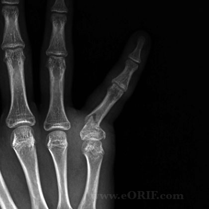
|
synonyms: phalanx base fracture
Phalangeal Base Fracture ICD-10
A- initial encounter for closed fracture
B- initial encounter for open fracture
D- subsequent encounter for fracture with routine healing
G- subsequent encounter for fracture with delayed healing
K- subsequent encounter for fracture with nonunion
P- subsequent encounter for fracture with malunion
S- sequela
Phalangeal Base Fracture ICD-9
- unspecified = 816.00 (closed), 816.10(open)
- proximal or middle phalanx = 816.01(closed), 816.11(open)
- distal phalanx = 816.02(closed), 816.12(open)
- multiple sites = 816.03(closed), 816.13(open)
Phalangeal Base Fracture Etiology / Epidemiology / Natural History
Phalangeal Base Fracture Anatomy
Phalangeal Base Fracture Clinical Evaluation
- evaluate finger cascade with flexion. Any overlaps of injured digits indicates need for reduction +/- fixation.
Phalangeal Base Fracture Xray
- P/A and lateral views of affected finger
- 30-45 degree suppinated or pronated views
Phalangeal Base Fracture Classification / Treatment
- Undisplaced, displaced unicondylar fx involving <25% of articular surface without joint subluxation/deformity: buddy taping
- Displaced unicondylar: Closed reduction under c-arm guidance, generally requires pointed reduction forceps. Cannulated reduction forceps facilitates K-wire placement. ORIF if closed reduction fails (Tan V, JOT 2005;19:518)
- Displaced Bicondylar: almost always unstable. CRPP. Generally joint is reduced and rixed with transverse k-wires placed parallel to joint surface. Metaphysis is then reduced and secured to the diaphysis with two K-wires inserted from the tip of each condyle proximally into the medullary canal distally. Also consider ORIF if closed recuction fails (Tan V, JOT 2005;19:518)
- Open fracture: consider mini-external fixation. (Freeland AE, CORR, 1987;214:93)
Phalangeal Base Fracture CRPP Technique
- Belsky MR, J Hand Surg 1984;9Am:725
- 0.045 (1.1mm) K-wires generally used. Consider 0.062in(1.6mm) for larger bones(metacarpal); 0.035in(0.9mm) for smaller bones (pediatric fx).
- Closed reduction under c-arm guidance.
- one or more intramedullary or two crossed K-wires.
- K-wires must not cross at the fracture site.
- K-wires are left in until fracture callus in visible on xray. Usually 3-4 weeks
Phalangeal Base Fracture ORIF Technique
- Mid-axial incision centered over fx
Phalangeal Base Fracture Associated Injuries
- Adjacent phalanx or metacarpal fractures
- Carpal fractures
- fight bite (high risk of injection)
- Phalangeal Shaft Fracture
- Phalangeal Neck Fracture
- Distal Phalanx Fracture
Phalangeal Base Fracture Complications
- Flexion Contracture: see Hogan CJ, JAAOS 2006;14:524.
- Loss of reduction
- Delayed union
- Malunion
- Tendon adhesion / stiffness
- Cold intolerance
- Numbness
- Abnormal nail growth
- Nerve or vascular injury
Phalangeal Base Fracture Follow-up
- Weekly follow-up until fracture callus is seen on xrays is indicated to ensure reduction is maintained
- Advance to passive ROM / Buddy taping as soon as fracture allows.
Phalangeal Base Fracture Review References
- Freeland AE, Geissler WB, Weiss AP, Operative Treatment of Common Displaced and Unstable Fractures of the Hand, JBJS 2001;83A:928-945
- Freeland AE, Orbay JL. ORIF of the Tubular Bones of the Hand In: Stickland JW, Grahma TJ, editor. Hand 2nd ed. Master Techniques in orthopedic surgery. Philadeplhia: Lippincott-Raven; 2004. p 3
|


