Peroneal Tendon Dislocation S86.399A 726.79


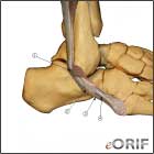
Peroneal tendon anatomy image

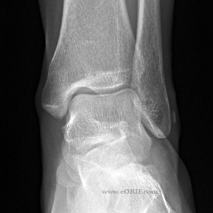
Fleck sign 

synonyms: peroneal tendon instability

Peroneal Tendon Instability ICD-10

Peroneal Tendon Instability ICD-9

  • Peroneal tendinitis ICD-9 = 726.79

Peroneal Tendon Instability Etiology / Epidemiology / Natural History

  • often accompanied by avulsion of the posterior osteochondral ridge of the distal fibula seen on plain xray
  • mechanism of injury (acute dorsiflexion and violent reflex peroneal musculature contraction) precipitates an avulsion of the superior peroneal retinaculum, allowing dislocation ot the peroneal tendons anteriorly.
  • Common in soccer and skiing.

Peroneal Tendon Instability Anatomy

  • Superior peroneal retinaculum: primary retaining structure for the perneal tendons. Origin: posterior margin of the distal 1-2cm of the fibula. Travels posteriorly to the lateral calcaneus with extensions into the paratenon of the Achilles tendon. Most commonly avulses from the fibula often with a small fleck of bone.  (Maffuli N, AJSM 2006;34:986).
  • Deficient posterior distal fibular groove may contibute to peroneal instability.  Approximately 25% of people have a flattened or convex peroneal groove.
  • Hindfoot varus alignment is a predisposition to peroneal tendon injury.
  • Os peroneum is a sesamoid bone present in 10% to 20% of people located along the peroneus longus near the peroneal groove of the cuboid. (Sobel M, Foot Ankle Int 1994;15:112)
  • Anatomic varients in peroneal tendon sheath: low-lying peroneal brevis muscle belly; anomalous peroneal quartus tendon.

Peroneal Tendon Instability Clinical Evaluation

  • Lateral ankle pain, swelling and ecchymosis.
  • Tenderness posterioly along the peroneal tendons.
  • Pain and weakness with active ankle dorsiflexion and external rotation.
  • Lateral tenderness, no medial tenderness.
  • May complain of popping in the posterolateral ankle with push off.
  • Ankle Circumduction Test:

Peroneal Tendon Instability Xray / Diagnositc Tests

  • A/P, Lateral and Mortise views of the ankle are generally normal. May demonstrate a "fleck" sign: small avulsion fracture of the lateral malleolus which is pathognomonic for acute dislocation of the peroneal tendons and indicates Grade III injury to the superior peroneal retinaculum.
  • MRI: demonstrates peroneus brevis +/- peroneus longes dislocated anterolateral to the lateral malleolus with disruption of the peroneal retinaculum.
  • Dynamic ultrasound: may be indicated to evaluated for pseudosubluxation of the peroneal tendons.

Peroneal Tendon Instability Classification / Treatment

  • Surgical exploration with reduction of tendons and repair of superior peroneal retinaculum +/- deepening of the retromalleolar groove(axially ream the fibula to remove cancellous bone and then impact the posterior cortex in the groove with a bone tamp; or trapdoor osteotomy of posterior fiblula).  Must also correct any varus hindfoot malalignment. If >50% of peroneal tendon cross-section is torn or attenuated consider peroneal tendon tenodesis (suture longus and brevis together). (Krause JO, Foot Ankle Int 1998;19:271).  (see AAOS The Athlete's Ankle Volume 1 for technique videos)
  • Pseudosubluxaiton: no subluxation for the retrofibular groove with dorsiflexion-eversion maneuver but with painful snapping symptoms related to the peroneus longus snapping through or over the peroneus brevis. Treatment = repair of tendon tears with peroneal groove deepening. (Raikin SM)
  • Poor outcome with non-op treatment. Consider bracing or taping for in season athletes with definitive surgical treatment after season ends. 

Peroneal Tendon Instability Associated Injuries / Differential Diagnosis

  • PER ankle fracture (Weber type C)
  • SER ankle fracture (Weber type B)
  • Proximal fibula fx (Maisonneuve).
  • Ankle dislocation.
  • Peroneal Tendon instability
  • 5th Metatarsal fracture
  • Calcaneal fracture
  • Ankle Instability

Peroneal Tendon Instability Complications

  • Recurrent instability
  • Continued pain
  • Stiffness
  • CRPS

Peroneal Tendon Instability Follow-up Care

  • Outcomes of peroneal tendon tear operative treatment:  90% return to employment at the same level. 58% scar tenderness; 54% lateral ankle swelling; 46% successfully return to sports.(Steel MW, Foot Ankle Int 2007;28:49)

Peroneal Tendon Instability Review References

  • Philbin TM, JAAOS 2009;17:306
  • Murr S: Dislocation of the peroneal tendon with marginal fracture of the lateral malleolus.  J Bone Joint Surg 1965;43B:563-565.
  • Eckert WR, Davis EA Jr: Acute rupture of the peroneal retinaculum.  J Bone Joint Surg 1976;58A:670-672.
  • Zoellner G, JBJS 1979;61A:292