 |
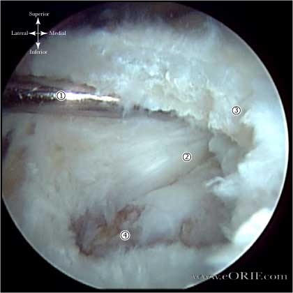
Cresent shaped
- Minimal retraction, excellent mobility.
- Generally repaired directly to bone.
|
 |
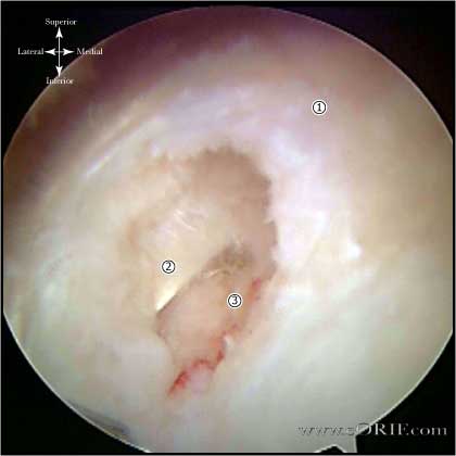
U-shaped
- similar to cresent tears but with retraction to the glenoid rim.
- Even after releases the apex of the tear is difficult to mobilize.
- Generally repaired with margin convergence followed by repair of remaining free margin to bone.
- Supraspinatus-- small U-shaped tear
- Long head of biceps tendon (normal)
- Humeral Head
Arthroscopic view from beach chair position
|
| |
L-shaped
- similar to U-shaped tears, except one leaf is more mobile than the other.
- Generally repaired by side-to-side suturing / margin convergence followed by repair of remaining free margin down to bone.
|
 |
Massive/contracted
- no mobility from medial to lateral or anterior to posterior.
- Require mobilization / interval slide techniques for repair. Anterior interval slide: release the interval between the supraspinatus and rotator interval using a cutting instrument introduced through a lateral portal. The release is oriented toward the base of the coracoid. Posterior interval slide: clear the scapular spine of its surrounding subacromial fibroadipose tissue so that it can be clearly seen through a lateral viewing portal. Place traction sutures in the infraspinatus and supraspinatus and split the interval between them. Avoid injury to the suprascapular nerve by lifting the soft tissue away from the bone when cutting, and stopping the release when the fat pad at the base of the scapular spine is visible.
-
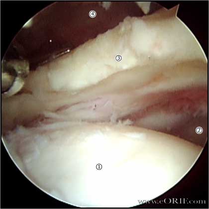
- Humeral head
- Glenoid
- Supraspinatus tendon
- Undersurface of acromion
|
| |
Partial thickness
- May be managed non-operatively with PT.
- Shoulder arthroscopy is indicated for partial thickness tears that fail non-operative management.
- Consider transtendon repair of partial thickness tears that fail nonoperative managment.
- Tear <50% of the RTC thickness may be debrided.
- >50% can be expanded to full thickness tears and repaired.
- Articular sided partial thickness tears may do poorly with debridement alone.
- Throwing athletes (pitchers) have a high incidence of failure to return to sport after RTC repair. Consider debridement up to 75% partial thickness tears with repair after career has ended.
|
 |
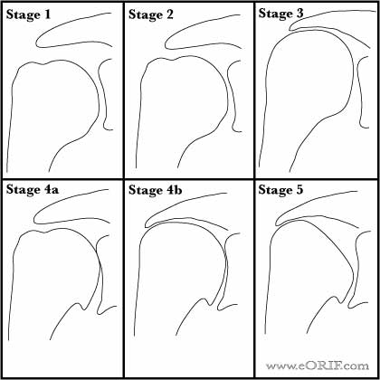
Hamada Classification of Arthritis in chronic rotator cuff tears (Hamada K, CORR 1990;254:92).
- Stage 1: Acromiohumeral interval greater than 6 mm.
- Stage 2: Acromiohumeral interval less than 7 mm.
- Stage 3: Acromiohumeral interval less than 7 mm with acetabulization of acromion.
- Stage 4a: Acromiohumeral interval less than 7 mm with glenohumeral arthritis without acetabulization.
- Stage 4b: Acromiohumeral interval less than 7 mm with acetabulization and glenohumeral arthritis.
- Stage 5: Acromiohumeral interval less than 7 mm with osteonecrosis of humeral head.
|
| |
Goutallier Classification of Muscle Atrophy (Goutallier D, CORR 1994;304:78).
- Stage 0=completely normal muscle.
- Stage 1=muscle contains some fatty streaks.
- Stage 2=fatty infiltration is important, but there is still more muscle than fat.
- Stage 3=there is as much fat as muscle.
- Stage 4=more fat than muscle is present.
- Degeneration is grade at the tip of the coracoid process and at the inferior margin of the glenoid and the values are averaged to determine the stage.
|
| |
Postoperative cuff integrity Classification
- Type I = sufficient thickness compared with normal cuff with homogenously low intensity on each image
- Type II = sufficient thickness compared with normal cuff associated with partial high intensity area
- Type III = insufficient thickness with less than half the thickness when compared with normal cuff, but without discontinuity, suggesting a partial-thickness delaminated tear
- Type IV = presence of a minor discontinuity in only 1 or 2 slices on both oblique coronal and sagittal images, suggesting a small full-thickness tear
- Type V: presence of a major discontinuity observed in more than 2 slices on both oblique coronal and sagittal images, suggesting a medium or large full-thickness tear.
- Sugaya H, Arthroscopy 2005;21:1307
|


