|


|
synonyms:shoulder dislocation, anterior shoulder instability,
Anterior GH Instability ICD-10
Anterior GH Instability ICD-9
- 718.31 Recurrent shoulder dislocation
- 831.01 Closed anterior dislocation of the shoulder
Anterior GH Instability Etiology / Epidemiology / Natural History
- Athletes: instability may occur with repetitive external rotation with the arm in abduction (apprehension position).
- May result from a traumatic dislocation with a Bankart lesion, bony-Bankart lesion, ALPSA, HAGHL, repetitive microtrauma with stretching of the anteroinferior capsulolabral complex or generalized ligamentous laxity with subtle subluxation and functional impairment.
Anterior GH Instability Anatomy
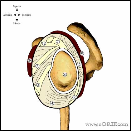
- Glenohumeral stability is dependent on static (labrum, glenohumeral osseous morphology, GH capsular ligaments) and dynamic (RTC, deltoid, biceps, scapular musculature, "concavity compression") restraints.
- Inferior glenohumeral ligament (consist of anterior inferior GH ligamant, axillary pouch and posterior inferior GH ligament) is the most important for resisting anterior translation in the abducted arm.
- RTC produces concavity-compression by creating a net force vector which directs the humeral head into the glenoid. The scapular stabilizers affect this net force by positioning the glenoid. 10º of scapular protraction significantly increases the strain on the anterior band of the IGHLC (Weiser WM, AJSM 1999;27:801).
- Deltoid acts as an anterior stabilizer in the abduction and externally rotated position (Kido T, AJSM 2003;31:399).
- Pectoralis Major forces increase anterior directed forces in the apprehension position (Labriola JE, JSES 2005;14:32S).
- 13% of patients have anatomic variants in the anterosuperior labrum (cordlike middle glenohumeral ligament, sublabral foramen, complete absence of labral tissue. (Rao AG, JBJS 2003;85A:653).
- 50% of anterior instability patients will have at least a small bony Bankart lesion (Sugaya H, JBJS 2003;85:878). 80% of bony Bankart lesions occur between the 2:30 and 4:20 positions (Saito H, AJSM 2005;33:889).
- Patient with recurrent anterior instability have impaired relflexive muscle activation in the apprehension position. Pec and biceps activation is suppressed; Subscap, supraspinatus and infraspinatus is increased (Myers JB, AJSM 2004;32:1013).
- Proprioception is impaired in patients with instability and is restored with surgical repair (Potzl W, AJSM 2004;32:425).
- 1-cm capsular plication = @ 20º loss of motion (Metcalf MH, JSES 2001;10:532).
- Glenoid is 23-30mm from anterior to posterior. 6-10mm bone defect = 25%/requires bone restoration. <3-4mm =15%/arthroscopic repair.
- See also Shoulder anatomy.
Anterior GH Instability Clinical Evaluation
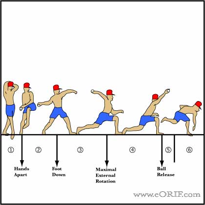
- Pain or instability sensation with the arm in abduction and external rotation. Shoulder feels like it will slide out the front in the late cocking phase of throwing.
- Throwers note a sensation of the shoulder sliding out the front during the late cocking phase of throwing. May be related to "dead arm syndrome" with transient sudden, sharp pain associated with loss of ball control and numbness.
- Anterior dislocations: prominence of the humeral head anterior, medial and inferior to the shoulder joint with a hollow region beneath the lateral deltoid. Loss of normal deltoid contour with prominent anterior acromion. Arm is held in an abducted, sligthly externally rotated position.
- Apprehension Test
- Relocation Test
- Load and shift
- Sulcus sign
- Evaluate for axillary nerve function. Humeral head may compress the axillary nerve.
- Evaluate Cervical Spine.
- Assessments of generalized ligamentous laxity: ability to touch ipsilateral forearm with the thumb, place palms of hands on floor with knees locked, elbow/knee/MCP hyperextension, patellar instability.
- Must rule out psychological problems/ voluntary dislocators!
- Assess for recurrence risk factors: uncontrolled seizures (epilepsy), alcoholism, drug dependancy, propensity for falls, high-load abduction and external rotation activities (kayaking).
- See also Shoulder Physical Exam.
Anterior GH Instability Xray / Diagnositc Tests
- A/P and Lateral view in the plane of the scapula, and axillary view. Generally normal.
- West Point view: patient prone with arm in 90° abduction and neutral rotation. Xray beam is directed 25° posterior to the horizontal plane and 25° medial to the vertical plane. Useful for evaluating the anterior glenoid rim / bony bankart lesions.
- Garth (apical oblique) view: provides view of glenoid bone loss and Hill-Sachs lesions.
- Hill-Sachs lesion: impression fracture of the posterolateral aspect of the humeral head, produced by contact with the anteroinferior glenoid when dislocated. Hill-Sachs lesion is demonstrated on plain AP radiograph in internal rotation or Stryker notch view.
- CT scan is best to evaluate bony anatomy and should be considered for the recurrent dislocator suspected of having a large Hill-Sachs or bony Bankart lesion.
- Glenoid Bone loss evaluation (Bigliani LU, AJSM 1998;26:41)
-Type I: displaced avulsion fracture
-Type II: malunited avulsion fracture
-Type IIIA: erosion of glenoid; less than 25%
-Type IIIB: erosion of glenoid; greater then 25%
- MRI arthrogram (gadolinium): the anterior and posterior labrum are best seen on axial images and appear as dark triangular structures. Bankart lesions appear as a loss of the normal triangular shape or contrast material may extend between the labrum and glenoid( acute injuries.) Chronic injuries may scar down to the glenoid and be difficult to see by MRI.
Anterior GH Instability Classification / Treatment
- Physical therapy focused on strenghtening program especially aimed at the dynamic glenohumeral and scapular stabilizers. Avoid provacative position for 8 weeks followed by gradually return to sport specific activities. Anterior Instability Rehab Protocol.
- Operative treatment: anterior plication / Bankart repair. generally reserved for failure of non-operative treatment. Open and arthroscopic techniques have similar outcomes (Bottoni CR, AJSM 2006;34:1730), (Fabbriciani C, Arthroscopy 2004;20:456).
- Acute Dislocation: see Shoulder Dislocation.
- Chronic Anterior Instability with Hills Sachs lesion: Lesions representing >30% of the articular surface (determined on pre-op CT) generally require treatment. Treatment options: humeral head allograft (Gerber C, JBJS 1996;78A:376), Remplissage (Wolf EM, Arthrosopy 2004;20(suppl1):e14,)
- Chronic Anterior Instability with Glenoid deficiency: Glenoid deficiency greater than 13.5% generally requires treatment (bone grafting vs coracoid transfer). Treatment options = Latarjet procedure (Allain J, JBJS 1998;80:841), (Burkart S, AJSM), vs Distal tibial allograft. Other options:Tricortical iliac crest great (Warner JJ, AJSM 2006:34:205), Open anterior soft tissue stabilization [large defects loose @7ºER] (Pagnani M, JBJS 2008;36A:1805).
- Failed Laterjet: Consider hardware removal and distal tibial allograft. (Provencher, MT, ASES2018)
- Evaluate patients over 45y/o for RTC tear.
Anterior GH Instability Associated Injuries / Differential Diagnosis
- Hills Sachs lesion
- Posterior Glenohumeral Instability
- Internal Impingement
- RTC Tear
- Posterior Capular contracture
- SLAP Lesion
- Subacromial Impingement
- HAGL Lesion
- Suprascapular nerve entrapment(Cummins CA JBJS-AM 2000;82:415-424).
- Quadrilateral space syndrome(Cahill BR J Hand Surg-AM 1983;8:65-69).
- Early osteoarthritis
- Tumor
Anterior GH Instability Complications
- Recurrent instability
- Pain
- Hardware failure / Anchor pull-out
- Infections
- Stiffness
- CRPS
- Nerve injury: Axillary nerve, Brachial plexus
- Fluid Extravasation:
- Chondrolysis: though to be related to heat from electo cautery or radiofrequency probes used during capsular release or capsular shrinkage.
- Hematoma
- Chondral Injury / arthritis
- Instrument failure
- Weakness
Anterior GH Instability Follow-up Care
Anterior GH Instability Review References
|


