Achilles Tendon Repair Case

Achilles Approach Anatomy

37 y/o female sustained Achilles tendon rupture playing tennis. Underwent repair 5 days after injury.

Achilles Approach Anatomy

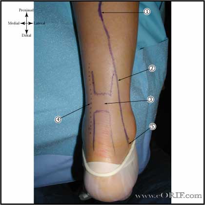
  1. Sural nerve crosses near the midline at the level of the musculotendinous junction of the achilles @9.8 cm from the calcaneus.
  2. Sural nerve
  3. Palpable defect in Achilles tendon generally in avascular zone 3-6cm proximal to calcaneal insertion.
  4. Incision along medial border of the Achilles tendon.
  5. Sural nerve is 18.8 mm from the lateral border of the Achilles tendon at the level of insertion of the Achilles into the calcaneus.


Achilles Approach Anatomy
  • Pre-op antibiotics.
  • Prone, all bony prominences well padded.
  • Thigh tourniquet.
  • Prep and drape in standard sterile fashion.
  • Posteromedial incision (medial to the medial border of the Achilles). Dissect down to paratenon (deep crural fascia), without making large skin flaps.
  • Paratenon incised longitudinally.
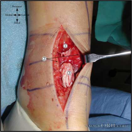
  • Hematoma evacuated, tendon ends debrided.
  1. Proximal stump, overlying fatty tissue and hematoma has not been debrided yet.
  2. Paratenon.
  3. Distal Stump.
Achilles repair picture
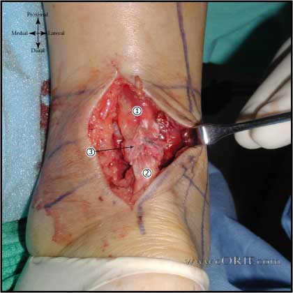
  • Repair performed with 2 #2 nonabsorbable sutures (Orthocord) placed using a modified-Bunnell suture technique. Sutures were tied within the tendon to prevent bulky suture mass.
  • Reinforced with a vertical locking circumferential 0-Vicryl suture.
  1. Proxmial stump
  2. Repair area, note bulky suture knots are within tendon mass and overwrapped with circumferential 0-Vicryl suture.
  3. Distal stump.
achilles tendon repair picture
  • Paratenon repaired with 2-0 absorbable suture.
  • Sub-Q closed with inverted interrupted 2-0 Vicryl.
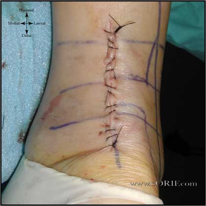
  • Skin closed with running 3-0 Nylon suture placed in baseball stitch configuration.
  • Placed in below-knee bulky compressive dressing with posterior plaster splint with foot in gravity equinus postion.