 |
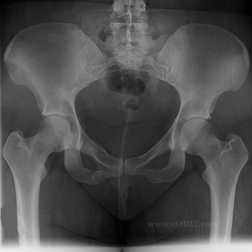
A/P Pelvis
- Helpful for: Hip arthritis, Pelvic fracture, acetabular fracture, femoral neck fracture, IT Fx, AVN, Adult DDH, etc
- Evaluate: joint space narrowing, osteophytes, cysts, asses leg lengths based on lesser troch levels. Pubic symphysis should line up with sacrum if pt and film are properly oriented.
- Lateral Center-edge angle of Wiberg
Position: supine with both legs internally rotated 15°.
Beam: aimed perpendicular to table/plate, centered 1" distal to ASIS.
|
 |
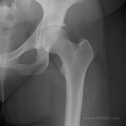
A/P Hip
- Helpful for: Hip arthritis, Pelvic fracture, acetabular fracture, femoral neck fracture, IT Fx, AVN, etc
- Evaluate: joint space narrowing, osteophytes, cysts,
Position: supine with both legs internally rotated 15°.
Beam: aimed perpendicular to table/plate, centered on femoral neck.
|
| |
Cross Table Lateral Hip
- Helpful for: Hip arthritis, Pelvic fracture, acetabular fracture, femoral neck fracture, IT Fx, AVN, etc
- Evaluate: joint space narrowing, osteophytes, cysts, fracture
Position: supine with unaffected hip and knee flexed 90° out of the way.
Beam: aimed perpendicular to long axis of femoral neck, centered on femoral neck.
|
| |
Frogleg Lateral Hip
- Helpful for: Hip arthritis, DDH, SCFE, AVN, etc
- Evaluate: joint space narrowing, osteophytes, cysts, fracture, femoral head coverage, hip joint congruity
Position: supine with hips flexed and abducted, knees flexed with soles of feet touching.
Beam: aimed perpendicular to table/plate, centered on pubic symphysis.
|
 |
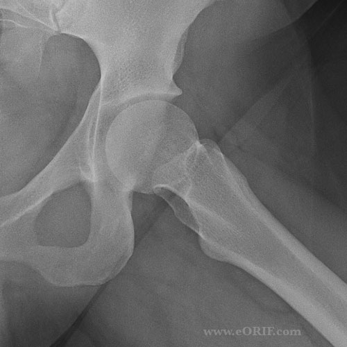
Lateral Hip
- Helpful for: Hip arthritis, DDH, SCFE, AVN, femoral neck fracture,etc
- Evaluate: joint space narrowing, osteophytes, cysts, fracture, femoral head coverage, hip joint congruity
Position: supine rotated posteriorly toward affected side. Hip is flexed 45° and abducted 45°.
Beam: aimed perpendicular to table/plate, centered on femoral neck.
|
 |
False Profile View
Lequesne and de Seze view
- Helpful for: DDH,
- Evaluate: anterior coverage of the femoral head
- Measeure ventral inclination angle. Normal >25º.
Position: Patient standing with the affected hip on the cassette, the ipsilateral foot parallel to the cassette and the pelvis rotated 65° from the plane of the cassette.
Beam: centered on the femoral head perpendicular to the cassette
Reference: Garbuz DS, CORR 2004;418:18, Lequense M, Rev Rhum Mal Osteoartic 1961;28:643
|


