|


|
synonyms: Distal third clavicle fracture, lateral clavicle fracture, distal clavicle fx
Distal Clavicle Fracture ICD-10
Distal Clavicle Fracture ICD-9
- 810.03 (closed fracture of acromial end clavicle)
- 810.13 (open fracture of acromial end clavicle)
Distal Clavicle Fracture Etiology / Epidemiology / Natural History
- 15% of clavicle fractures
- Often unstable and prone to malunions and nonunion.
- Typically take longer to heal the mid shaft clavicle fractures with an average time to union of approximately 3 months.
Distal Clavicle Fracture Anatomy
- AC ligaments stabilize the joint in the anteroposterior direction.5,6 The superior AC ligament is the stongest of the AC ligaments. Its fibers blend with the fibers of the deltoid and trapezius muscles. Distal clavicle resection, which is part of many described treatment methods, leads to anteroposterior instability due to loss of the AC ligaments.7 The posterior and superior AC ligaments should be preserved during distal clavicle resection to prevent posterior instability. (Klimkiewicz JJ, JSES 1999;8:119)
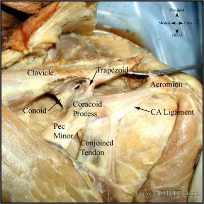
- Coracoclavicular ligament is made up of the trapezoid and conoid ligament. It is the prime suspensory ligament of the upper extremity.8
- Trapezoid ligament in men has a mean length of 1.61 +/- 0.52 and a width of 1.58 +/-0.69 cm.5 Orgin = coracoid process, anterior and lateral to the attachment of the conoid ligament. Insertion = a rough line on the undersurface of the clavicle extending anteriorly and laterally from the conoid tubercle.
- Conoid ligament in men has a mean length of 1.225 +/- 0.658 cm and width of 0.737 +/-0.148 cm.5 Origin = the posteromedial side of the base of the coracoid process. Insertion = conoid tubercle on the posterior undersurface of the clavicle. The conoid tubercle is located at the apex of the posterior clavicular curve, which is at the junction of the lateral third of the flattened clavicle with the medial two thirds of the triangular-shaped shaft.
Distal Clavicle Fracture Clinical Evaluation
- Pain over clavicle, crepitus and motion at fracture site. Often obvious deformity.
- Evaluate skin over fracture site for tenting, inpending skin compromise
- Ptosis of affected shoulder.
- NV exam of upper extremity indicated; rule out axillary artery/vein injury.
- Auscultate chest to rule out associated pneumothorax.
Distal Clavicle Fracture Xray / Diagnositc Tests
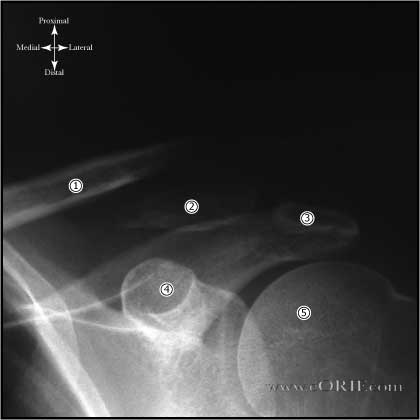
- A/P view of clavicle and 30° cephalic tilt view of clavicle.
- Apical oblique (Grashe with 20 cephalad)
- Abduction Lordotic (after ORIF)
- Serendipity view (helps with A/P displacement)
- Axillary
- Zanca 15 degree apical oblique (AC joint)
- Consider chest xray (including both clavicles) if there is significant deformity (shortening) or if concerned for pneumothorax or rib Fracture's.
- CT with 3-D reconstruction is helpful for segmental fractures or for medial clavicle fractures which may be missed on Chest xrays.
Distal Clavicle Fracture Classification / Treatment
- (later sub divided be Neer and Rockwood)
- Type I: distal to the coracoclavicular ligaments. Coracoclavicular ligaments remain intact, displacement uncommon. Treatment = sling.
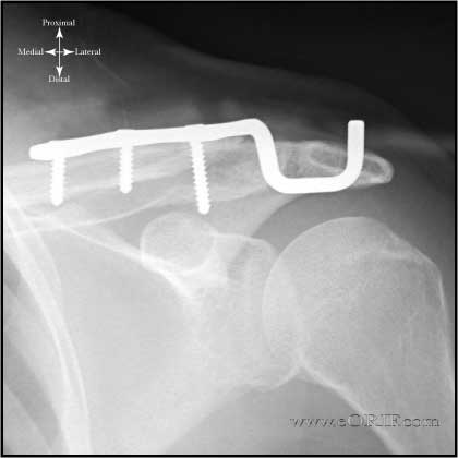
- Type II: medial to (type IIA) or between (type IIB) the coracoclavicular ligaments. Medial fragment lfrequently displaces superiorly. Nonunion is frequent. Treatment = ORIF. Consider hook-plate fixation (Haider SG, JSES 2006;15:419) (Meda PV, Injury 2006;37:277). Consider non-absorbable suture fixation. (Levy O, JSES 2003;12:24).
- Type III: intra-articular, frequently without ligament disruption. Generally little or no displacement. Frequently missed or mis-diagnosed as acromioclavicular joint injuries. May lead to Acromioclavicular Arthritis.
- ORIF fixation options: distal radius T-plate (Kalamaras, JSES 2007;17:60), hook-plate fixation (Haider SG, JSES 2006;15:419) (Meda PV, Injury 2006;37:277),
- Pediatric distal clavicle fractures: generally through the distal physis and adjacent metaphysis. The acromioclavicular joint is rarely dislocated; inherently stable and heal well with conservative treatment.
Distal Clavicle Fracture Associated Injuries / Differential Diagnosis
- Pneuomothorax=3% (Rowe, CORR 58;29:1968)
- Brachial Plexus=rare (usually traction injury)
- Subclavian artery=rare(intimal tear)
Distal Clavicle Fracture Complications
- Nonunion: more common with Type II Distal Clavicle fractures.
-Treatment = Clavicle Fracture ORIF +/- bone grafting.
- Malunion:
- CA ligament ossification
- Infection
- Neurovascular Injury
Distal Clavicle Fracture Follow-up Care
- Post-op: sling for comfort, no overhead motion. Immediate pendelum ROM exercises.
- 10-14 Days: Wound check, sutures removed. Start PT for gentle ROM exercises. No resistive exercises/activities. Sling as needed for comfort.
- 6 weeks: Xrays, if union is evident begin strengthening and resistive exercises. No contant athletics.
- 3 months: Repeat xrays. If pt is painfree and union is obvious pt may return to sport. If no signs of union, consider bone stimulator, see Nonunion.
Distal Clavicle Fracture Review References
- Nuber GW, JAAOS, 1997;5:11
|


