|

|
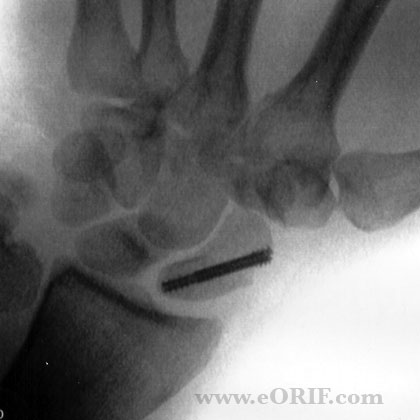
synonyms:scaphoid percutaeous screw fixation, scaphoid fracture fixation, scaphoid ORIF, scaphoid repair, carpal navicular fracture ORIF, carpal navicular percutaneous screw fixation
Scahoid Fracture CPT
Percutaneous Scaphoid Fixation Indications
- Nondisplaced scaphoid waist fracture
- Acute unstable scaphoid waist fractures which can be reduced closed.
- Proximal pole fractures
- Fibrous nonunions without avascular necrosis
Percutaneous Scaphoid Fixation Contraindications
- Fractures in which adequate reduction cannot be achieved via a closed manner.
- Scaphoid nonunions with sclerosis, cystic changes, pseudarthrosis, avascular necrosis, and humpback deformities.
Percutaneous Scaphoid Fixation Alternatives
- Short arm thumb spica cast / long arm thumb spica cast
- ORIF
Percutaneous Scaphoid Fixation Planning / Special Considerations
- Screws must be placed in the central third of both poles of the scaphoid for greatest stability, and rapidity of fracture healing.
- Proximal pole fractures should be approached dorsally.
- Waist fractures may be approached dorsally or volarly.
- Distal fractures are best approached volarly.
- Displaced fractures may be reduced via: volar traction-assisted approach; the dorsal minimal incision approach, with manual reduction as the guidewire is advanced; or the dorsal approach with arthroscopy-assisted reduction.
- Proximal approach: distal aiming point is the center of the scaphotrapezial joint or the base of the thumb. Starting point = just radial to the ulnar proximal corner of the scaphoid at the insertion of the scapholunate ligament
- Distal approach: proximal aiming point is the ulnar proximal corner of the scaphoid at the insertion of the scapholunate ligament.
- Scaphoid Case card.
- Arthrex headless compression screw
- Acumend Acutrak headless compression screw
- Depuy Synthes Headless compression screw
- Stryker Cannulated compression screw
Percutaneous Scaphoid Fixation Technique
- Sign operative site.
- Pre-operative antibiotics, +/- regional block.
- General endotracheal anesthesia
- Supine position. All bony prominences well padded.
- C-arm positioned on opposite side of the patient. Pronation and suppination of the forearm allows complete 180° fluorscopic visualization of the scaphoid.
- Perfrom fluoroscopic exam of the wrist. Evaluate for occult carpal fracture, great arc injury, scapolunate instability.
- Prep and drape in standard sterile fashion.
- Wrist suspended from the thumb finger trap.
- Insert 12-gauge needle into the scaphotrapezial joint and then pass guidewire down the needle.
- Double-checking guidewire position with multiple radiographic views.
- Ensure screw is fully buried beneath the articular cartilage. Screw lenght should be 4mm shorter than the measured length of the scaphoid.
- Irrigate.
- Close in layers.
Percutaneous Scaphoid Fixation Complications
- Nonunion
- Malunion
- Scaphoid subsidence or shortening with secondary screw penetration
- Neurovascular injury (cutaneous nerve, radial artery)
- Incorrect placement of a fixation screw
- Failure to recognize concomitant injuries.
Percutaneous Scaphoid Fixation Follow-up care
- Post-op: Place in volar thumb spica splint.
- 7-10 Days: If rigid fixation in good bone was achieved start controlled motion program with a removable splint and range-of-motion and gripping exercises.
- 6 Weeks: Consider CT to determine when union has occurred. Unprotected activity is not allowed until bridging bone is seen
- 3 Months: Consider bone stimulator if union not confimed
- 6 Months: assess ROM
- 1Yr: follow-up xrays, assess outcome
Percutaneous Scaphoid Fixation Outcomes
- 100% union at an average of twelve weeks. Fractures treated earlier heal more quickly than those treated later. (Slade JF, Gutow AP, Geissler WB: JBJS 2002; 84A (suppl 20): 21-36).
- Screw fixation: 7wks to union, 8 weeks return to work. Cast: 12 weeks to union, 15 weeks return to work. No significant difference in wrist ROM or grip strength at 2 years. (Bond CD, JBJS 2001; 83A: 483-488).
Percutaneous Scaphoid Fixation Review References
|

