|


|
synonyms: PIP Dislocation(PIP=Proximal interphalangeal joint)
PIP Fracture / Dislocation ICD-10
A- initial encounter
D- subsequent encounter
S- sequela
PIP Fracture / Dislocation ICD-9
- 834.02(closed IP joint dislocation)
- 834.12(open IP joint dislocation)
PIP Fracture / Dislocation Etiology / Epidemiology / Natural History
- usually direct blow and an axial load to tip of digit such as a ball in sporting event
PIP Anatomy
- 85% of motion for grasping objects occurs at PIP joint
- PIP joint has the largest arc of motion (120 degrees) of three joints in each digit
PIP Fracture / Dislocation Clincal Evaluation
- Generally have obvious pain and deformity of the PIP joint.
- Document neurovascular status of the finger before and after any reduction.
PIP Fracture / Dislocation Xray
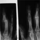
- A/P and lateral of affected digit.
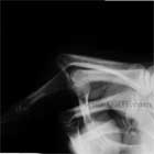
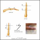
- True lateral xray should show a congruent joint surface with two parallel surfaces. “V” sign indicates incomplete reduction.
PIP Fracture / Dislocation Classification / Treatment
- PIP Dislocation without fracture
-Doral dislocation=volar plate is ruptured, usually at distal attachment on middle phalanx. May be clinically inconsequential avulsion fragment. RX: dorsal block splint at 20-30 degrees for 1 weeks followed by AROM with buddy taping.
-Volar dislocation= rare; rotatory compression on semi-flexion middle phalanx; usually unilateral collateral ligament disruption and partial volar plate avulsion, completely volar may rupture central slip of extensor mechanism. Test ability to maintain full extension after reduction. If stable begin mobilization. If unstable consider immobilization of the PIP in extension or with surgical repair of the central slip. Central slip insufficiency leads to formation of a boutonniere deformity.
-After reduction, these injuries are frequently immobilized for too long of a time period and in too much flexion. As a result, the ruptured volar plate contracts, scars, and shortens as it heals. After the injury, it is important to mobilize the joint as soon as the patient is comfortable. The patient will sometimes require intensive physical therapy to get the proximal interphalangeal joint extended again. Redislocation or subluxation, arthritis, and collateral ligament instability are much less common.
- PIP Dislocation with Prox phalanx fracture
-Inspect for rotational deformity
-may involve one or both condyles
-RX: immobilization with frequent f/u, CRPP, ORIF, Ex fx. At least two implants are required for rotational stability. Dorsal approach(exposes both condyles risk ext mech scaring), Midlateral approach(risks NV bundle)
-loss of motion common. Mean PIP motion at 3 yr f/u=71 degrees. (Weiss A, J Hand Surg 1993;18A;594)
- PIP Dislocation with Dorsal lip Middle Phalanx fracture
-hyperflexion injury
-usually <25% of articular surface
-can be avulsion of central slip
-nondisplaced=splint in extension. Mobilize at 3-4 wks with continued splinting for 6-8 wks. At 8wks start aggressive mobilization
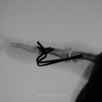
-Displaced=ORIF to avoid boutonneire deformity. Dorsal approach, mini-screw vs tension band vs k-wires.
- PIP Dislocation with Palmar lip Middle Phalanx fracture
-more common
-hyperextension(usually <30% with little comminution) or axial load injury(articular comminution)
-<30% articular surface fracture is typically stable after reduction. Treatment = short period of immobilization followed by buddy taping with ROM exercises.
->50% of articular surface fracture is unstable. Treatment = ORIF vs CRPP/extension block pinning.
->60% articular cartilage comminuted and impacted: hemi-hamate arthroplasty with bone graft indicated.
- PIP Joint Pilon Fracture
-comminution at base of middle phalanx (pilon fx)is best treated with indirect reduction through dynamic splinting or the use of specially designed PIP joint external fixators. (Badia A, J Hand Surg 2005;30A:154) (Stern PJ, J Hand Surg 16A:844;1991), (Krakauer J, CORR 327:29;1996)
PIP Fracture / Dislocation Associated Injuries / Differential Diagnosis
- MCP dislocation
- Phalangeal Base Fracture
- Phalangeal Neck Fracture
- Distal Phalanx Fracture
- Mallet finger
PIP Fracture / Dislocation Complications
- Tendon adhesion / stiffness
- Flexion Contracture: see Hogan CJ, JAAOS 2006;14:524.
- Infection
- Malunion
- Nonunion
- Nerve or vascular injury
- Loss of reduction
- Delayed union
PIP Fracture / Dislocation Fracture Follow-up
- Post-op /Initial: Splint. Elevation.
- 7-10 Days: xray to ensure reduction is maintained. Continued splint, activity modifications. Immobilize as few joints as necessary.
- 6 Weeks: Remove k-wires, wean from splint use as soon as callus is visible on xray. Continue activity modifications. Agressive ROM.
- 3 Months: Resume full activities. Assess ROM. May require flexor/extensor tendon tenolysis to regain motion.
- 1Yr: assess outcomes / follow-up xrays.
PIP Fracture / Dislocation Review References
- Rockwood and Greens
- Greens Hand Surgery
|


