|


|
synonyms: Golfers elbow, medial epicondylitis, common flexor tendon tear, common flexor tenosynovitis, handball player's elbow
Medial Epicondylitis ICD-10
Medial Epicondylitis ICD-9
- 726.31 (medial epicondylitis)
Medial Epicondylitis Etiology / Epidemiology / Natural History
- Pain in the medial aspect of the elbow due to fibrillary degeneration of collagen, angiofibroblastic hyperplasia , vascular granulation tissue, tendinous necrosis and secondary inflammation of the flexor-pronator muscle mass.
- golf, rowing, baseball pitching, javelin, tennis, bricklaying, hammering, typing, textile production
- 4th to 5thdecade
- male=female
- 7-20 times less common than lateral epicondylitis
Medial Epicondylitis Anatomy
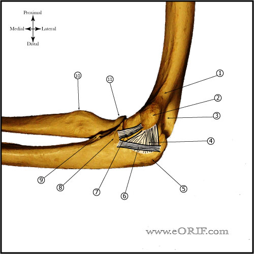
- medial epicondyle=flexor pronator muscle mass origin
- from prox to distal = pronator teres, FCR, palmaris longus, FDS, FCU
- pronator teres and FCR, most commonly involved, both arise form medial supracondylar ridge
- throwing – peak angular velocity and valgus stress occur primarily during acceleration phase(0 forward velocity to ball release)
Medial Epicondylitis Clinical Evaluation
- Pain along the medial elbow.
- Pain exacerbated by resisted forearm pronation and wrist flexion.
- Tenderness over the origin of the flexor-pronator muscle mass.
- Normal elbow/wrist ROM
- Evaluate for Cubital Tunnel Syndrome(holding elbow maximally flexed with wrist extended for 3 minutes produces pain and numbness, Tinel's at elbow)
- Evaluate for Posterolateral Rotatory Instability
- Note
Medial Epicondylitis Xray / Diagnositc Tests
Medial Epicondylitis Classification / Treatment
- Nonsurgical treatment = avoid offending activity, NSAIDs, physical therapy, counterforce bracing,
- Physical therapy =wrist flexor and forearm pronator stretching and progressive isometric exercises advancing to eccentric/concentric exercises as tolerated
- Corticosteriod injection: benefits remain controversial. Double-blind studies have indicated no long term benefit. Risks / complications include: fat atrophy, depigmentation of skin, disruption of muscle origin, post-injection flare, facial flushing, iatrogenic infection. (Stahl S, JBJS 1997;79Am:1648)
- Surgical Treatment Indications: failure of nonoperative management for a minimum of 6 to 12 months. Debridement of pathologic tissue, release of the flexor carpi radialis - pronator teres origin, and/or repair of the flexor carpi radialis - pronator teres origin. Concomitant ulnar neuropathy may have worse outcomes following surgery.
Medial Epicondylitis Surgical Technique
- (Vangsness CT Jr, JBJS Br 1991;73:409-411)
- supine, arm board, tourniquet, pre-op antibiotic
- 8cm longitudinal incision centered over medial epicondyle
- Common flexor origin reflected
- Ensure medial collateral ligament is preserved and ulnar nerve is protected.
- Pathologic tissue on undersurface of the flexor pronator mass in excised.
- Medial epicondyle is debrided/drilled to a bleeding bone bed.
- Common flexor pronator origin reattached with suture
- Closure
- Posterior splint
Medial Epicondylitis Associated Injuries / Differential Diagnosis
Medial Epicondylitis Complications
- continued pain
- infection
- ulnar nerve palsy
- stiffness
- instability
Medial Epicondylitis Follow-up Care
- Splint removed at 7-10 days. Gentle elbow ROM started. Hand wrist ROM encouraged.
- Resisted wrist flexion/pronation started at 6 weeks
- Return to activity at 4 months post-op
- Generally anticipate return to prior activity levels after surgery without loss of flexor/pronator strength.
- Excellent/Good results obtained in 97% of pts. 86% had no limitations(Vangsness CT Jr, JBJS Br 1991;73:409-411)
Medial Epicondylitis Review References
|


