| |
Lachman Test
Lachman (Torg JS, Am J Sports Med. 1976;4:84-93)
best test for ACL laxity. Knee placed in 20-30 degress of flexion, the femur is stabilized, and an anteriorly directed force applied to proximal calf. Compare to uninjured side. Grade 1+ = 1-5mm increased translation; 2+= 6-10mm ; 3+=>10mm.
|
| |
Pivot Shift
Confirms complete ACL tear. Based on very early flexion causing anterior subluxation of the tibia that is reduced with further flexion (20-40 degrees) due to the posterior pull of the iliotibial tract. Relocation event graded as 0(absent), 1+ (pivot glide), 2+(pivot shift), 3+(momentary locking) (Galway HR, JBJS 1972;54Br:763)
|
 |
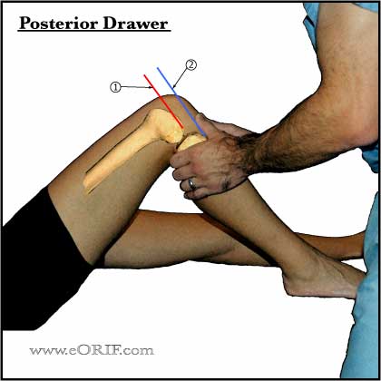
Posterior Drawer
- Most accurate test for PCL tear.
- Patient in supine postion with knee flexed 90°.
- Palpate the medial tibial plateau and appreciate its position relative to the medial femoral condyle.
- Apply a posteriorly directed force to proximal tibia.
- Note the change in the distance (the step-off) from the medial tibial plateau and medial femoral condyle.
Figure: Normal Posterior Drawer
Step-off between medial femoral condyle and medial tibial plateau is maintained when posteriorly directed force is applied to proximal tibia.
- Medial femoral condyle
- Medial tibial plateau
|
|

|
Posterior Drawer
- Grade I = palpable but diminished stepoff (0-5mm).
- Grade II = lost their stepoff, but the medial tibial plateau cannot be pushed beyond the medial femoral condyle (5-10mm).
- Grade III = complete PCL injuries stepoff is lost, and the tibia can be pushed beyond the medial femoral condyle (>10mm) and has an obvious positive posterior sag.
Figure: Grade II Posterior Drawer
Step-off between medial femoral condyle and medial tibial plateau is lost (5-10mm of subluxation) when posteriorly directed force is applied to proximal tibia.
- Medial femoral condyle
- Medial tibial plateau
|
| |
Quadriceps Active Test |
| |
Dial Test(Tibial External Rotation):
- Prone postion. Measure thigh-foot angle with external rotation stress applied both at 30° and 90°. Compare to normal side. External rotation of the tibia >10° compared to normal side indicates posterolateral corner injury. Increased ER at 30° but not at 90° indicates isolated posterolateral corner injury. Increased ER at both 30° and 90° indicates combined PLC, PCL injury. (Bleday RM, Arthroscopy 1998;14:489-94) (Larsen MW, J Knee Surg 2005;18:146-50).
|
| |
Valgus Laxity
- Indicates MCL injury or posteromedial corner injury.
|
| |
Varus Laxity
|
| |
McMurray test
- patient lying supine with the hip and knee flexed 90°. Apply axial compression while ER and IR the leg.
- Reproduction of pain +/- clicking indicates meniscal tear.
|
| |
Posterolateral external rotation test
- Posterior tanslation and external rotation forces are applied to the knee at 30° of flexion. Posterolateral subluxation of the proximal tibia indicates PLC injury.
|
| |
External rotation recurvatum test
- Lift the patients extended leg by the great toe and observe for any side to side differences in hyperextension, varus and tibial external rotation. (Hughston JC, CORR 1980;147:82-7).
|
| |
Posterolateral drawer test
- (Hughston JC, CORR 1980;147:82-7), adduction stress test, dynamic posterior shift test, Reverse pivot shift test.
|
| |
Anterior drawer test -
anterior force applied at 90 degrees of flexion. Least reliable.
|
| |
Ege's Test
- Place feet 8-10 inches appart with feet pointed outward (medial meniscus) or inward (lateral meniscus.
- Patient squates down with feet flat of the floor. Pain or a click when the knee approaches 90 indicates meniscal tear.
- 71% accuracy, 67% sensitivity, 81% specificity for medial meniscal lesions (Akseki D, Arthroscopy, 2004;20:951).
|
| |
Apley Compression Test |


