|
 
|
Synonyms: IT fracture, intertroch fracture, hip fracture
Intertrochanteric Femur Fx ICD-10
A- initial encounter for closed fracture
B- initial encounter for open fracture type I or II
C- initial encounter for open fracture type IIIA, IIIB, or IIIC
D- subsequent encounter for closed fracture with routine healing
E- subsequent encounter for open fracture type I or II with routine healing
F- subsequent encounter for open fracture type IIIA, IIIB, or IIIC with routine healing
G- subsequent encounter for closed fracture with delayed healing
H- subsequent encounter for open fracture type I or II with delayed healing
J- subsequent encounter for open fracture type IIIA, IIIB, or IIIC with delayed healing
K- subsequent encounter for closed fracture with nonunion
M- subsequent encounter for open fracture type I or II with nonunion
N- subsequent encounter for open fracture type IIIA, IIIB, or IIIC with nonunion
P- subsequent encounter for closed fracture with malunion
Q- subsequent encounter for open fracture type I or II with malunion
R- subsequent encounter for open fracture type IIIA, IIIB, or IIIC with malunion
S- sequela
Intertrochanteric Femur Fx ICD-9
- 820.21(closed)
- 820.31(open)
Intertrochanteric Femur Fx Etiology / Epidemiology / Natural History
- 5% initial hopstial mortality, 12%-30% one-year mortality for all hip fracture patients.
- Mortality Risk Factors: age >85, lack of independent ADL's prior to fracture, poor nutrition, pre-op ASA class3 or 4, CHF, pneumonia. Stongest predictor = dementia. (Morrision RS, JAMA 2000;284:47), (Richmond J, JOT 2003;17:52).
- Preoperative risk factors associated with postoperative mortality: chronic renal failure, congestive heart failure, chronic obstructive pulmonary disease, age of older than 70 years. (Bhattacharyya T, J Bone Joint Surg Am. 2002 Apr;84-A(4):562-72)
- Elderly hip fractures are associated with low-energy falls and osteroporsis. Hip fractures in young patients are associated with high-energy trauma (MVA, fall from height).
- Non-op treatment/delayed surgical treatment = decubiti, UTI, joint contractures, pneumonia, DVT/PE, high mortality rate.
Intertrochanteric Femur Fx Anatomy
- IT fractures occur between the greater and lesser trochanters.
- iliopsoas inserts on the lesser trochanter and flexes, externally rotates, and shortens the limb.
- Gluteus medius and minimus insert on the greater trochanter and produce varus deformity at the fracture site and shorten the limb.
- Short external rotators often remain intact, externaly rotating the proximal fragment
- Hip adductors insert distal to the fracture causing adduction and varus deformity.
Intertrochanteric Femur Fx Clinical Evaluation
- Lower extremity is shortened and externally rotated with displaced fractures.
- Tender eccymotic hip.
- Pain with any hip ROM.
- Document lower extremity NV exam.
- Document any pre-existing sacral or heel decubitus ulcer.
Intertrochanteric Femur Fx Xray
- AP pelvis, cross-table lateral hip, AP hip. AP in 15° of internal rotation are best to demonstrate non-displaced fractures.
- MRI: indicated for suspected hip fracture not seen on xray. Consider bone scan if MRI is not available. Bone scans may not be positive for up to 3 days after fracture
Intertrochanteric Femur Fx Classification / Treatment
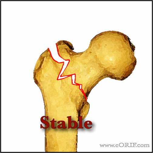
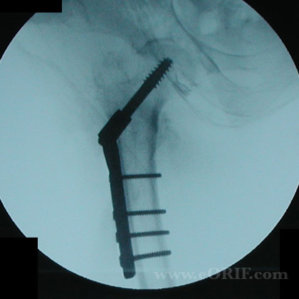
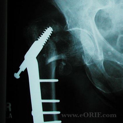
- Stable fracture (posteromedial cortex intact/minimal comminution): 2-hole DHS. Tip-apex distance shoulde be equal to or less than 25mm (tip apex distance = the sum of the distance from the tip of the lag screw to the apex of the femoral head on the AP and lateral Xrays) (Baumgaertner MR, JBJS 1995; 77:1058).
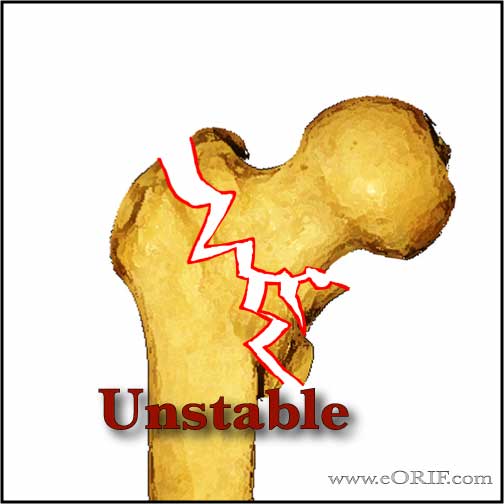
- Unstable fracture (posteromedial cortex comminuted, lateral cortex disruption, subtrochanteric extension): IMHS. Generally with long unlocked nail, unless there is subtrochanteric extention. Distal locking indicated for fractures with subtroch extention. Increased failure/complications rate.
- Reverse oblique IT Fxs (5% of all IT/subtrochanteric fxs): Sliding hip screw is associated with many complications and failures. 95 degree fixed-angle device or intramedullary hip screw with distal locking are indicated. (Haidukewych JBJS 2001;83A:643)
- Nonambulatory with minimal pain / medically unstable: Nonoperative treatment (early mobilization is key. Will have varus shortened malunion / nonunion)
- Patholigc fracture: long intramedullary nail.
- Preoperative skin traction in patients with hip fractures does not provide significant pain relief, as compared with pillow placement under the injured extremity (Rosen JE, JOT 2001;15:81), (Anderson GH, JBJS 1993;75Br:794) (Parker MJ, Cochrane Database of Systematic Reviews 2006;3:1).
- Surgical delay greater than 4 days is associated with increased mortality, UTI, decubitus ulcers, DVT and pneumonia. ((Moran CG, JBJS 2005;87A:483), (Rodriquea_Fernandez P, CORR, 2011)
- Regional anaesthesia: decreeased 1-month mortality and DVT compared to general anesthesia. GErnal anesthesia has reduced OR time. (Urwin SC, Br J Anaesh, 2000;84:450), (Luger TJ, Osteo Int'l 2010;21:555).
- Elderly pts with fragility fractures should be evaluated for osteoporosis.
- Orthofix pertrochanteric Fixator (Moroni A, JBJS-AM 2005;87:753-9).
- Gotfried PCCP www.orthofix.com, percutaneous plating system.
- AO Classification (AO Classification)
- IMHS Technique
- DHS Technique
Intertrochanteric Femur Fx Associated Injuries / Differential Diagnosis
Intertrochanteric Femur Fx Complications
- Screw cut-out: associated with tip-apex distance >25mm, increasing age of the patient, an unstable fracture, a poor reduction, & use of a high-angle (150deg) side-plate
- Loss of fixation
- Nonunion
- Malunion (malrotation)
- Osteonecrosis of the femoral head
- Femoral shaft fracture (IMHS)
- Painful hardware
Intertrochanteric Femur Fx Follow-up
- After hip fracture the vast majority of patients require assistance in performing ADLs. Only 20% to 35% of patients who were independent in ADLs before fracture will regain their prefracture ADL independence. Factors predictive of recovery of function in ADL are younger age, absence of dementia or delirium in nondemented patients, and a strong social network. (Koval KJ, JAAOS 1994;2:141)
- 41% maintain their prefracture ambulatory ability at a minimum followup of 1 year; 40% remain ambulatory but become more dependent on assistive devices; 12% previous community ambulators became household ambulators; 8% become nonfunctional ambulators (Koval KJ,Clin Orthop Relat Res 1995;310:150)
- Elderly pts with fragility fractures should be evaluated for Osteoporosis.
Intertrochanteric Femur Fx References
- Rockwood and Greens
- Kellam JF, Fischer TJ, Tornetta P III, Bosse MJ, Harris MB(eds): OKU Trauma 2. Rosemont, IL, AAOS, 2000, pp 125-131.
|


