|
|
synonyms:DIP Fusion, DIP arthrodesis, distal interphalangeal joint fusion, distal interphalangeal joint arthrodesis
DIP Fusion CPT
DIP Fusion Indications
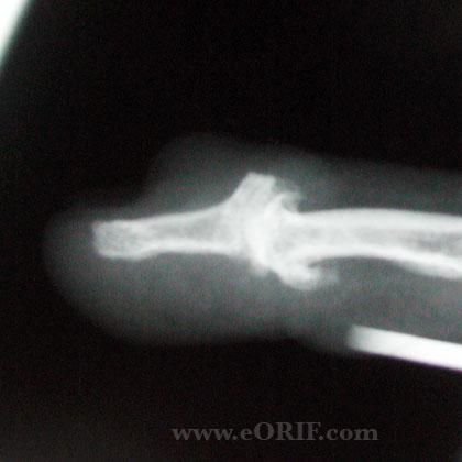
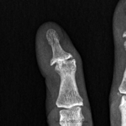
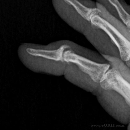
- DIP joint pain, deformity and funtional loss secondary to arthritis(osteo/inflammatory/post-traumatic/post-infectious).
- Chronic tendon rupture
- Burns
DIP Fusion Contraindications
- Active infection
- Occupation or hobby which maintenance of DIP motion
DIP Fusion Alternatives
DIP Fusion Planning / Special Considerations
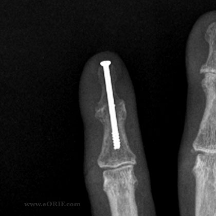
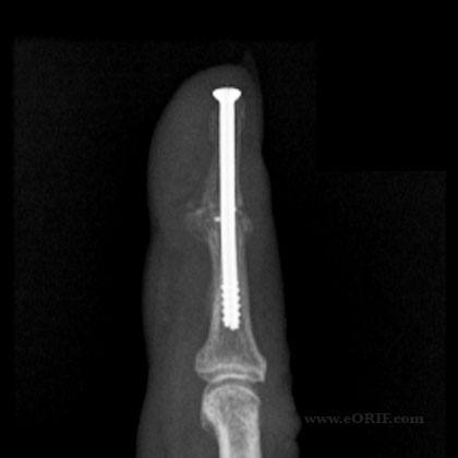
- Fixation options: K-wires, tension bands, intraosseous wiring, headless bone screws(Herbert, Acutrak, EBI VPC, Synthes; generally must be 3.0mm or small screw), plates
- DIP joint is generally fused in full extension.
- PIP joint IF, MF fused 15-30°.
- PIP joint RF, SF fused at 30-45° .
DIP Fusion Technique
- Pre-operative antibiotics, +/- regional block
- Digital block anesthesia +/- intravenous sedation.
- Supine position. All bony prominences well padded.
- Prep and drape in standard sterile fashion.
- Penrose drain used as a tourniquet.
- Dorsal H, transverse or Y-incision.
- Extensor mechanism is exposed and incised transversely exposeing the DIP joint.
- Release collateral ligaments if needed.
- Use small oscillatin saw to make the joint surfaces flat.
- Predrill middle phalanx centrally with a k-wire.
- Under c-arm guidance drill 0.062-inch double tipped K-wire from proximal to distal centrally in the distal phalanz.
- Reduce joint and drive K-wire into middle phalanx centrally.
- Verify placement with fluoroscopy.
- Drill per screw manufacture recommendations.
- Place appropriate screw.
- Verify reduction and screw placement with fluoroscopy.
- Irrigate.
- Close in layers.
DIP Fusion Complications
- Pin-tract infection.
- Deep infection/osteomyelitis.
- Painful hardware.
- Nonunion.
- Cold intolerance.
DIP Fusion Follow-up care
- Post-Op: bulky hand dressing with volar splint. Keep elevated.
- 10-14 Days: Wound check, Stack splint applied. Active and passive PIP ROM encouraged.
- 6 Weeks: review xrays for signs of union.
- 3 Months: return to full activities if pain free and union evident by xray.
DIP Fusion Outcomes
DIP Fusion Review References
- Greens Hand Surgery
- Master Techniques in Orthopaedic Surgery-The Hand
|