|
 

|
synonyms:
Congenital Radial Head Dislocation ICD-10
Congenital Radial Head Dislocation Etiology / Epidemiology / Natural History
- most common congenital anomaly of the elbow
- etiology unknown
- 47% anterior, 43%posterior, 10% lateral (Almquist EE, JBJS 51A:1118 1969).
- usually asymptomatic with minimal functional limitations
- often bilateral
- may become painful in adolescence or adulthood due to degenerative changes.
- 60% are associated with abnormalities, syndromes, mental retardation or scoliosis (Sachar K, Hand Clin 1998;14:39).
- Posterior congenital dislocations may have autosomal-dominant or X-linked recessive inheritance (Reichenbach H, Am J Med Genet 1995;55:101).
Congenital Radial Head Dislocation Anatomy
- Radial head is a secondary stabilizer to valgus stress, with the primary restraint being the medial collateral ligament. Pathologic valgus instability results if the radial head is resected when there is concomitant medial collateral ligament injury
- Studies of static loading across the elbow have suggested that as much as 60% of force is transmitted across the radiocapitellar articulation. (Morrey BF, JBJS 1988;70A:250).
- Force transmission across the radial head is greatest between zero and 30 degrees of flexion, decreases with increased flexion. Force transmission is greater when the forearm is pronated than when it is supinated (Morrey BF, JBJS 1988;70A;250).
Congenital Radial Head Dislocation Clinical Evaluation
- posterolateral elbow prominence, restricted elbow extension and forearm rotation, elbow popping,
- May have pain with activity.
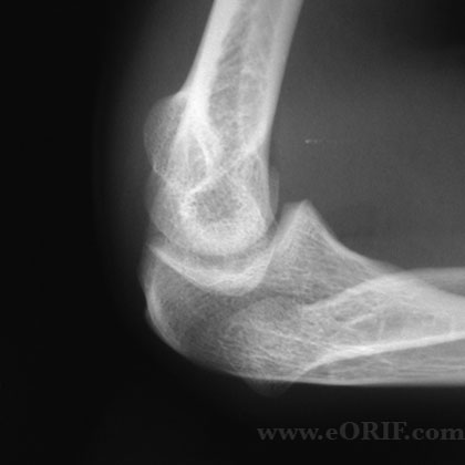
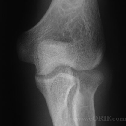
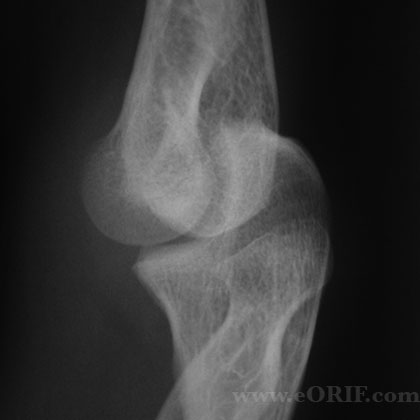
Congential Radial Head Dislocation Xray
- A/P, lateral and oblique elbow films indicated.
- radial head is dome shaped with no central depression, flattened or hypoplastic capitellum, mild bowing of the proximal ulna
Congenital Radial Head Dislocation Classification Treatment
- non-operative management strongly recommend
- may be treated by radial head resection if symptomatic after skeletal maturity.
Congenital Radial Head Dislocation Associated Anomalies
- 60% occur in association with a syndrome, strongly consider referral to geneticist
- congenital radioulnar synostosis
- Klinefelter’s
- Cornelia de Lange
- Ehlers-Danlos
- Nail-patella syndrome
- Silver's syndrome
Congenital Radial Head Dislocation Complications
- if radial head resected before skeletal maturity=prox radius regrowth, post-op radioulnar synostosis, cubitus valgus deformity.
- PIN palsy, synostosis, proximal migration of radius, valgus instability, cubitus valgus, wrist pain.
Congenital Radial Head Dislocation Follow-up Care
- Asymptomatic patients can be as active in sports as their function allows. May consider limiting contact sports.
Congenital Radial Head Dislocation Review References
- Sachar K, Hand Clin 1998;14:39
- Miura T. J Hand Surg 1990;15Br:477
- Mardam-Bey T, J Hand Surg 1979;4Am:316– 20
a |


