Calcaneus Avulsion Fracture S92.009A 825.0


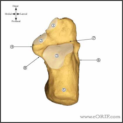
Calcaneus anatomy image

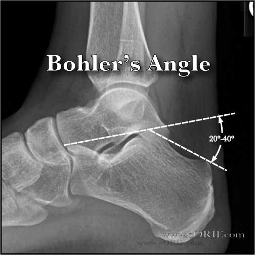
Bohler's angle calcaneus xray

Gissane's angle calcaneus xray


synonyms: os calcis fracture, calcaneous avulsion  fracture

Calcaneus Avulsion Fracture ICD-10

Calcaneus Avulsion Fracture ICD-9

  • 825.0(closed)
  • 825.1(open)

Calcaneus Avulsion Fracture Etiology / Epidemiology / Natural History

  • uncommon

Calcaneus Avulsion Fracture Anatomy

  • Transmits body weight to ground, strong lever arm for calf muscles
  • Main weight beaing surface is the posterior facet
  • Anterior process: a saddle-shaped projection of bone at the superior aspect of the calcaneal body. Its inferior surface articulates with the cuboid. The bifurcate ligament inserts on the anterior process and connects the cuboid and navicular bones. Serves as the origin of part of the extensor digitorum brevis.
  • Peripheral structures of the calcaneus include the sustentaculum tali, the peroneal tubercle, and the medial and lateral calcaneal tubercles.
  • Tibial nerve crossses the calcaneous below the sustentaculum tali medially and may be injured with displaced fractures causing incomplete loss of sensation on the plantar surface of the foot.

Calcaneus Avulsion Fracture Clinical Evaluation

  • Document smoking history, diabetes, PVD (increased risk of wound complications).
  • Document plantar foot sensation (risk of tibial nerve injury with incomplete loss of sensation on the plantar surface of the foot.)

Calcaneus Avulsion Fracture Xray / Diagnositc Tests

  • A/P, Lateral, and Mortise Ankle and A/P, Oblique, Lateral foot xrays indicated. 
  • Harris view(axial view): demonstrates degree of lateral migration of the tuberosity.
  • Broden's view-demonstrates articular surface of posterior facet-pt supine, cassette under leg/ankle, foot in neutral flexion with leg internally rotated 30-40, beam centered over lateral malleolus; xrays taken at 40, 30, 20, 10 degrees toward head of patient.  Shows posterior facet as it moves from posterior to anterior. (Bruden Acta Radiol 31:85;1949). Generally get CT instead.
  • Bohlers angle(on lateral xray) usually between 20°-40°; formed by lines drawn from the highest point of the anterior process of the calcaneous to the highest point of the posterior facet and a line drawn tangential to the superior edge of the tuberosity.  Decrease indicates posterior facet has collapsed.
  • Gissane's Angle(on lateral xray) seen directly inferior to the lateral process of the talus, formed by lines along lateral border of posterior facet and anteriorly to the beak of the calcaneous. Usually between 95° and 105°.

Calcaneus Avulsion Fracture Classification / Treatment

  • Immediate open reduction and internal fixation.  Delayed treatment risks necrosis of the overlying soft tissue. 
  • Nonsurgical management leaves a large void which is unlikely to fill with bone; causes Achilles weakness and risks skin breakdown.

Calcaneus Avulsion Fracture Associated Injuries / Differential Diagnosis

Calcaneus Avulsion Fracture Complications

  • Wound breakdown 
  • Heel deformity/malunion
  • Peroneal tendon impingement / instability
  • Malunion
  • Heel pad pain
  • Heel exostoses
  • CRPS
  • Infection / Osteomyelitis

Calcaneus Avulsion Fracture Follow-up Care

  • Bulky jones dressing with posterior splint post-operative. Must keep foot elevated for first 36-72 hours to decrease incidence of wound healing problems.
  • 7-10 day f/u:calcaneous xrays, SLC, NWB. Consider cam-walker with gentle ROM exercises.
  • 4 wk follow-up: calcaneous xrays, compression stocking, cam-walker boot, gentle ankle/subtalar ROM exercises, NWB. Sub-talar motion = figure-of-eight motion, drawing alphabet with the great toe.
  • 3 months f/u: calcaneous xrays, 25% WB, progress to full WB over 1-2 weeks. Start PT for ROM, gait training.
  • 6 months f/u: calcaneous xrays, evaluate function outcome, ROM.
  • 1 year f/u: calcaneous xrays, evaluate function outcome, ROM.

Calcaneus Avulsion Fracture Review References

  • Beavis RC,. Avulsion fracture of the calcaneal tuberosity: a case report and literature review. Foot Ankle Int. 2008 Aug;29(8):863-6
  • Hess M,. Calcaneal avulsion fractures: complications from delayed treatment. Am J Emerg Med. 2008 Feb;26(2):254

°