|


|
For other areas of osteonecrosis see: Kienbock's Disease (lunate), Preiser's Disease (scaphoid), Osteonecrosis of the knee. Osteonecrosis of Humeral Head
synonyms: avascular necrosis, AVN, osteonecrosis
Osteonecrosis ICD-10
Osteonecrosis ICD-9:
- 733.42 (head of femur), 733.41(head of humerus), 733.44(talus), 733.43(medial femoral condyle), 733.40(unspecified), 733.49(other)
Osteonecrosis Etiology / Epidemiology / Natural History
- Most commonly adults 20-50yrs old, 60% bilateral, 10% of primary THA
- Most often occurs in hips. 15% other bones affected, humeral head, knee, talus, small bones of hands/feet.
- Develops after 10% of undisplaced femoral neck fx, 15-30% of displaced fx and 10% of hip dislocations.
- 70% due to EtOH or prolonged high dose steriods. Other risk factors: sickle cell, dysbarism, Gaucher's disease, trauma, high-dose radiation, thrombophilia, protein S & C defiencies, hypofibrinolysis.
- Thought to be due to altered circulating lipids and coagulation mechanisms.
- Marrow death occurs 6-12 hrs after ischemia, radiographic changes appear after 3 months, articular collapse within 6-12 months in 80% of patients with clinical AVN.
Osteonecrosis Anatomy
- Lateral epiphyseal artery, which is the terminal branch of the medial femoral circumflex artery of the profunda femoris circulation supplies the majority of the femoral head.(Trueta, JBJS 35B:442;1953).
- Blood supply to femoral neck=extracapsilar arterial ring at base of neck supplied by branches of lateral and medial femoral circumflex artery, ascending cervical branches of arterial ring on surface of the neck, arteries of the ligamentum teres.
- AVN most commonly involves the lateral and anterior femoral head.
Osteonecrosis Clinical Evaluation
- Groin pain exacerbated by hip internal rotation.
- Duchene sign = pt leans to the affected side while in stance phase of gait; indicates hip pathology.
- Patrick's test: Groin pain with forcing hip into figure-of-4 postion, indicates hip pathology
- Stinchfield test: resisted straight leg raise causes groin pain, indicates hip pathology
Osteonecrosis Xray / Diagnositc Tests
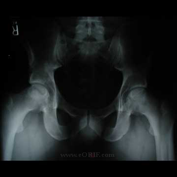
- AP pelvis, cross-table lateral hip, AP hip c leg internally rotated 15.
- 45 degree flexed knee A/P view best shows anterior superior femoral head lesion (90% of lesions)
- 90% of lesions are in anterior superior femoral head. Best viewed c 45° flexed knee A/P.
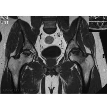
- MRI: If suspected but not seen or if only in one hip MRI is indicated. MRI sensitivity and specificiaty=95% for early AVN. Not good for estimating the extent of lesion.
- CT is the best technique to determine area of bone death.
- Bone scans are sensitive but had large number of false positives and have no advantage over MRI
- Evaluation should include lipid levels and possible screening for decreased levels of protein C and S and antithrombin III and increased levles of plasminogen activatoter inhibitor-1.
Osteonecrosis Classification / Treatment
ARCO Staging (Association for Research on Osseous Circulation) ARCO News 1992;4:41
- Stage 0-Bone biopsy demonstrates AVN. All other tests normal. Generally treated non-operatively
- Stage I-Bone scan or MRI positive. Lesions subdivided based on location (medial, central and lateral) and percentage of head involvement. Ia=<15% involvement; Ib=15-30%; Ic=>30%. Treatment: consider core decompression in young patients or Alendronate 70mg weekly (Lai KA, JBJS 2005;87A:2155).
- Stage II-Xray=osteoclerosis, cystic , osteopenia or mottled femoral head without collapse or acetabular involvement. Bone Scan/MF+RI=lesions subdivived based on location (medial, central and lateral) and percentage of head involvement. IIa=<15% involvement; IIb=15-30%; IIc=>30%. Treatment: core decompression for IIa sclerotic disease in young patients.
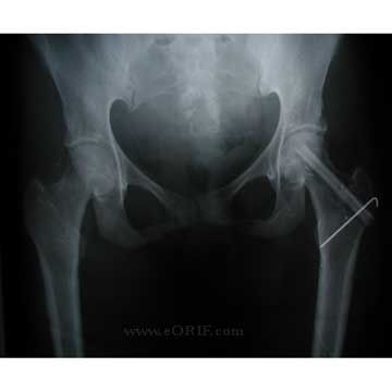
- Stage III-Xray=crescent sign; lesions subdivided based on location (medial, central and lateral) and percentage of head involvement. IIIa=<15% involvement or <2mm depression of head; IIIb=15-30% or 2-4mm depression; IIIc=>30% or >4mm depression. Treatment: Osteotomy vs vascularized fibula vs resurfacing vs THA.
- Stage IV-Xray=flattened particular surface, joint space narrowing, acetabular changes, osteophytes. Treatment: arthrodesis vs THA
- Ficat based on plain radiographs. Stage I=symptomatic hip,no xray change. Stage II, xray= patchy areas that are radiolucent and radiodense. Stage III=“crescent sign”=subchondral collapse. Stage IV=articular surface collapse.
- U of Pennslvania system (Steinberg). 0-normal MRI, XRAY, bone scan.1-abnormal MRI\bone scan .2-sclerotic\cystic changes on xray. 3-crescent sign. 4-flattened femoral head. 5-joint narrowing. 6-advanced DJD
Osteonecrosis Treatment Options
- Avoid chronic steriod use, correct lipid and coagulation abnormalities.
- conservative tx/ resticted weight bearing usually ineffective. May be indicated for very small asymptomatic lesions (ARCO stage I). (Cheng EY, JBJS 2004;86A:2594)
- electrical stimulation -some studies have shown benefit from pulsing electromagnetic fields applied externally. further study needed
- Alendronate 70mg weekly (Lai KA, JBJS 2005;87A:2155).
- Core Decompression - (CPT=27299: compare to 27071: or use s2325) removal of 8-10mm core of bone from femoral head and neck. Often provides prompt relief of pain. 64% achieve satisfactory clinical results compared to 23% for those treated non-surgically. prognosis is better the earlier the lesion is found and the smaller the lesion. Indicated for stage I or IIa disease. 6weeks protected weight-bearing post-op. (Bozic JBJS 1999;81A:200)
- Free vascularized fibular graft- 65% survival at 5years. (CPT=20955, 27170) (Aldridge JM, JBJS 2004 86A suppl 1;87) (Urbaniack JR, JBJS 1995;77A:681). 16.9% complications rate, 4.3% of complications require reoperation or chronic pain management (Gaskill TR, JBJS 2009;91A;1861)
- Osteotomy-Consider only for your symptomatic patients with Ficat Stage 2 disease. (Fuchs B, CORR 2003;412:84)
- Femoral Head Resurfacing: (CPT= )62.5% patient satisfaction, 75.9% 3 year survivorship (Adili A, CORR 2003;417:93)
- THA- If pt first encountered late with significant head collapse prophylactic measures are ineffective. generally use symtomatic treatment until reconstructive surgery indicated. Studies comparing unipolar and bipolar replacements to THA clearly favor THA.
Osteonecrosis Associated Injuries / Differential Diagnosis
Osteonecrosis Complications
- Femoral head collapse
- Arthritis
- Pain
- Free vascularized fibular graft complications = great-toe flexion contracture, ankle pain, nerve injury, infection, pin migration, HO, femoral fracture, DVT, Infection, hematoma, trochanteric bursitis (Gaskill TR, JBJS 2009;91A;1861)
Osteonecrosis Follow-up Care
- Dependent on stage and treatment.
Osteonecrosis Review References
|


