Ankle Arthroscopy 29891

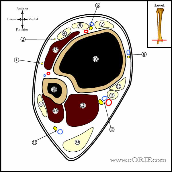
Ankle Cross section image

synonyms: ankle ATS, ankle scope, ankle arthroscopy

Ankle Scope CPT

29891 29897 
29894 29898
29895  29899

Ankle Scope Anatomy

  • anterolateral portal risks superficial peroneal nerve
  • anterocentral portal risks deep peroneal nerve or dorsalis pedis artery
  • anteromedial is just medial to the anterior tibial tendon, risks saphenous nerve
  • posterolateral portal risks sural nerve

Ankle Scope Indications

  • meniscoid lesions(fiborcartilaginous mass develops laterally after inversion ankle sprain causing chronic arterolateral ankle pain. 
  • synovitis
  • synovial chondromatosis
  • loose bodies
  • impingement(osteophytes)
  • OCD

Ankle Scope Contraindications

  • Infection

Ankle Scope Alternatives

  • Non-operative treatment
  • Open procedures

Ankle Scope Pre-op Planning / Special Considerations

  • Allows visualization of: transverse ligament, anterior tibiofibular ligament, deep deltoid ligament, deep portion of posterior tibiofibular ligament. Anterior talofibular ligament is poorly visualized.

Ankle Scope Technique

  • Pre-operative antibiotics, +/- regional block
  • anesthesia
  • supine position. All bony prominences well-padded.
  • Examination under anesthesia.
  • prep and drape standard sterile fashion
  • noninvasive ankle distractor
  • anatomic landmarks marked on skin (talus, medial mal, lateral mal, anterior tibial tendon, peroneous tertius, lateral Achilles tendon, subq course of superficial peroneal nerve dorsal intermediate branch ( invert foot and ankle, plantarflex lateral toes)), dorsalis pedis artery,
  • anteromedial portal site medial to anterior tibial tendon at the joint line localized with 18-gauge spinal needle
  • skin incised, subq tissues blunted dissected to level of capsule using fine hemostat (mosquito)
  • portal created with blunt obturator and cannula
  • anterolateral portal site determined with spinal needle lateral to peroneus tertius tendon
  • posterolateral portal created under arthroscopic visualization.  Spinal needle lateral to Achilles tendon (generally 2cm distal to anterior portals)
  • resect synovium
  • Anterior 8:deltoid ligament, medial gutter, medial talus, central talus, lateral talus, talofibular articulation, lateral gutter, anterior gutter
  • Posterior 3 from anterior portal: posteroinferior tibiofibular ligament, transverse tibiofibular ligament, capsular reflection of FHL, medial & lateral synovial recesses.
  • Posterior: medial gutter, medial central and lateral talus, talofibular articulation, lateral gutter, posterior gutter.
  • OCD lesions drilled with 0.062mm K-wire at 3mm interval to a depth of @ 10mm.
  • portal sites closed with 4-0 nylon,
  • bulky compressive dressing
  • well-padded posterior splint

Articular Cartilage Grading

  • A=smooth, intact but soft or ballotable
  • B=rough surface
  • C=fibrillations/fissures
  • D=Flap present or bone exposed
  • E=Loose, undisplaced fragment
  • F=Displaced fragment

Ankle Scope Complications

  • overall 9% complication rate (Ferkel Arthroscopy 1994 10:352)
  • neurologic: most common (primarily superficial branch of peroneal N, saphenous N or sural N. (Ferkel RD, Arthroscopy 1996;12:200).
  • vascular injury
  • superficial/deep infection
  • adhesions
  • fracture
  • ligament injur
  • incisional pain

Ankle Scope Follow-up care

Ankle Scope Outcomes

Ankle Scope Review References

  • Urguden M, Soyuncu Y, Ozdemir H, Sekban H, Akyildiz FF, Aydin AT. Arthroscopic treatment of anterolateral soft tissue impingement of the ankle: evaluation of factors affecting outcome. Arthroscopy. 2005 Mar;21(3):317-22.
  • Ferkel RD, Small HN, Gittins JE. Complications in foot and ankle arthroscopy. Clin Orthop Relat Res. 2001 Oct;(391):89-104.
  • Barber FA, Click J, Britt BT. Complications of ankle arthroscopy. Foot Ankle. 1990 Apr;10(5):263-6.
  • Feiwell, Foot Ankle 1993;14:142-147
  • °