 |
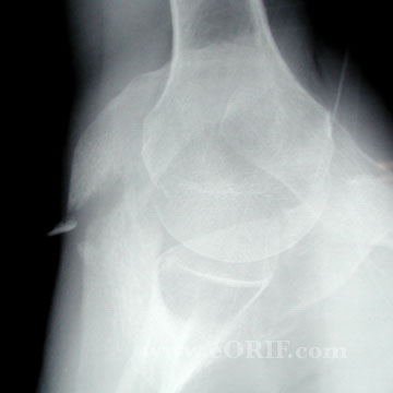
50y/o male sustained a displaced Type III acromion fracture in a fall onto shoulder. Axillary injury view shown. |
 |
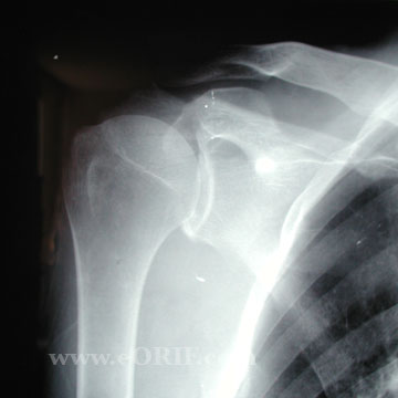
AP injury view of acromion fracture. |
 |
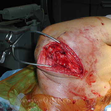
Underwent ORIF via a posterior Judet approach. Surgical exposure shown. Patients is in lateral decubitus position with the head to left in the image. |
 |
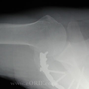
Reduction and fixation was performed with a Synthes 3.5mm pelvic reconstruction plate and non-locking cortical screws. |
 |
Follow up AP xray of acromion fracture. |
 |
Follow up axillary view of acromion fracture following ORIF. |
