Muscles of the Foot
| Muscle | Innervation | Origin | Insertion | Action | Nerve Root | |
| Dorsal layer | ||||||
| 1 | Extensor digitorum brevis(EDB) | Deep peroneal | superolateral calcaneus | base of proximal phalanges | extend | |
| First Plantar Layer | ||||||
| 1 | Abductor hallucis | med plantar | calcaneal tuberosity | base of great toe prox phalanx | abduct great toe | |
| 2 | Flexor digitorum brevus(FDB) | med plantar | calcaneal tuberosity | distal phalanges of 2nd-5th toes | flex toes | |
| 3 | Abductor digiti minimi | lat plantar | calcaneal tuberosity | base fo 5th toe | abduct small toe | |
| Second Plantar Layer | ||||||
| 1 | Quadratus plantae | lat plantar | med & lat calcaneus | FDL tendon | helps flex distal phalanges | |
| 2 | Lumbricals | med & lat plantar | FDL tendon | EDL tendon | flex MTP, extend IP | |
| 3 | FDL & FHL | tibial | tibia/fibula | distal phalanges of digits | flex toes/invert foot | |
| Third Plantar Layer | ||||||
| 1 | Flexor hallucis brevis(FHB) | med plantar | cuboid/lat cuneiform | proximal phalanx of great toe | flex great toe | |
| 2 | Adductor hallucis | lat plantar | oblique:2nd-4th metatarsals, Transverse:MTP | proximal phalanx of great toe lat | adduct great toe | |
| 3 | Flexor digiti minimi brevis(FDMB) | lat plantar | base of 5th metatarsal head | proximal phalanx of small toe | flex small toe | |
| Forth Plantar Layer | ||||||
| 1 | Dorsal interosseous(DIO) | lat plantar | metatarsal | dorsal extensors | abduct | |
| 2 | Plantar interosseous | lat plantar | 3rd-5th metatarsals | proximal phalanges medially | adduct toes | |
| 3 | (Peroneus longus and tibialis posterior | superficial peroneal/tiba | fibula/tiba | med cuneiform/navicular | everts/invert foot |
 |
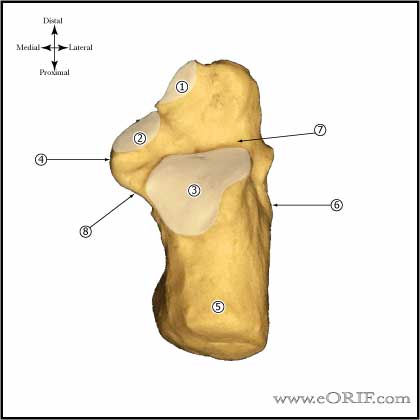
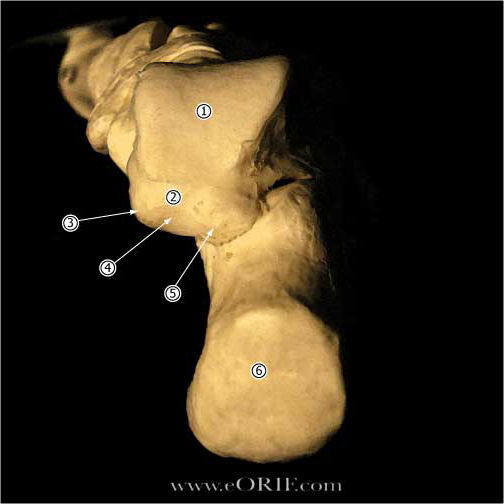
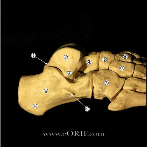
Calcaneous Anatomy |
| Talus Anterior portion of the talar dome is @2.4mm wider than the posterior portion, making the ankle much more stable in dorsiflexion than in plantarflexion. |
|
 |
|
| Spring ligament (calcaneonavicular) extends from the anterior aspect of the sustentaculum tali to the plantar medial surface of the navicular. Supports the plantar medial margin of the talar head. Two main components: superomedial calcaneonavicular ligament and inferior calcaneonavicular ligament. Superomedial calcaneonavicular ligament is commonly attenuated or torn in adult-acquired flatfoot deformity. | |


