|

|
synonyms: both bone forearm fracture, radius and ulna fracture
Pediatric Forearm Fracture ICD-10
A- initial encounter for closed fracture
B- initial encounter for open fracture type I or II
C- initial encounter for open fracture type IIIA, IIIB, or IIIC
D- subsequent encounter for closed fracture with routine healing
E- subsequent encounter for open fracture type I or II with routine healing
F- subsequent encounter for open fracture type IIIA, IIIB, or IIIC with routine healing
G- subsequent encounter for closed fracture with delayed healing
H- subsequent encounter for open fracture type I or II with delayed healing
J- subsequent encounter for open fracture type IIIA, IIIB, or IIIC with delayed healing
K- subsequent encounter for closed fracture with nonunion
M- subsequent encounter for open fracture type I or II with nonunion
N- subsequent encounter for open fracture type IIIA, IIIB, or IIIC with nonunion
P- subsequent encounter for closed fracture with malunion
Q- subsequent encounter for open fracture type I or II with malunion
R- subsequent encounter for open fracture type IIIA, IIIB, or IIIC with malunion
S- sequela
Pediatric Forearm Fracture ICD-9
- 813.23(fracture of the radius and ulna; closed)
- 813.33(fracture of the radius and ulna; open)
Pediatric Forearm Fracture Etiology / Epidemiology / Natural History
- Associated with tromplines, monkey bars, obesity (Davidon P, J Peadiatr Child Health 2003;39:664), poor milk intake (Gouldig A, J Am Diet Assoc 2004;104:250).
- Generally fall onto outstretched arm.
Pediatric Forearm Fracture Anatomy
- radial tuberosity and radial styloid should be on opposite sides of the radius.
- Ulnar styloid is on the opposite side of the ulna form the coronoid process.
Pediatric Forearm Fracture Clinical Evaluation
- Pain, swelling, deformity in forearm.
- Document neurovascular exam before and after any treatment.
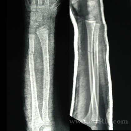
Pediatric Forearm Fracture Xray / Diagnositc Tests
- radial tuberosity and radial styloid should be on opposite sides of the radius.
- Ulnar styloid is on the opposite side of the ulna form the coronoid process.
Pediatric Forearm Fracture Classification / Treatment
- <10 y/o generally heal and remodel with no loss of rotation
- >10 y/o high risk of functional deficit, ORIF if unstable by closed means
- Acceptable reduction: complete displacement, angulation <10°(Price CT, JPO 1990;10:505-712): distal 1/3=20°, middle third=15°, proximal 1/3=10° for girls 8 years of age or younger and boys age 10 or younger
- acceptable reduction:<10 y/o angular correction of 1degree per month or 10degree per year, no rotational deformity, bayonet apposition with <1 cm shortening: >10yrs should be managed like adults(no deformity accepted)
- Reduction Analgesia: propofol/fentanyl (Godambe SA, Pediatrics 2003:112:116), ketamine/midazolam, axillary block (Kriwanek K:, JPO 2006:26:737), nitrous oxide with hematoma block (Luhmann JD, Pedicatrics 2006:118:e1078).
- Immobilize in LAC that incorporates thumb.
- Pediatric Forearm Fracture IM Nailing 25575 indicated for refractures with displacement, open fx, displaced proximal forearm fx, unstable fx, unacceptable alignment following CR, polytrauma
- ORIF with plates is generally not recommend in pediatric patients due to an increased risk of subsequent fracture at the end of the plates.
Pediatric Forearm Fracture Associated Injuries / Differential Diagnosis
Pediatric Forearm Fracture Complications
- poor results associated with proximal fxs, age >10, rotational malalignment, loss of radial bow
- Malunion=angulation >10, displacement >50%, malrotation, encroachment on interosseous membrane. Angulation of 20-30 degrees may be observed for 3 months for remodeling. Angulation >30 degrees corrective osteotomy is indicated. Restoration of motion is best if osteotomy is performed within 1year of injury. (Trousdale RT, JBJS Am 1995;77:894-902)
- Price CT, Scott DS, Kurzner ME, et al: Malunited forearm fractures in children. J Pediatr Orthop 1990;10:705-712.
- Kay S, Smith C, Oppenheim WL: Both-bone midshaft forearm fractures in children. J Pediatr Orthop 1986;6:306-310.
- refracture: greenstick fx’s have greatest risk of refracture. Strongly consider ORIF/flexible nails (Schwartz N, JBJS Br 1996;78:740-744)
- compartment syndrome
- delayed union
- Non-union (usually involves the ulna)
- infection
- superficial radial nerve palsy (generally transient)
- Growth arrest
Pediatric Forearm Fracture Follow-up Care
Pediatric Forearm Fracture Review References
- Huber RI, et al.: Flexible intramedullary nailing as fracture treatment in children. JPO, 1996;16:602-605.
- Flynn ICL 2002 51:355-60
- Flynn ICL 52:635-45
- CORR 402-245
|