|


|
synonyms: patellar fracture
Patella Fracture ICD-10
Patella Fracture ICD-9
- 822.0 (fracture of patella, closed)
- 822.1 (open)
Patella Fracture Etiology / Epidemiology / Natural History
Patella Fracture Anatomy
- Patella = sesamoid bone within the extensor mechanism of the knee.
- Bipartite patella ; present in @2% of patients.
Patella Fracture Clinical Evaluation
- Knee pain, effusion usually after a direct blow to the knee.
- Inability to extend the knee.
Patella Fracture Xray / Diagnositc Tests
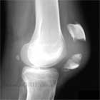
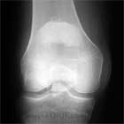
- A/P, lateral , Merchant or sunrise view of the knee. Fracture is generally best seen on lateral view.
- MRI: consider for patients with potential osteochondral fracture. Generally not needed.
- Intra-articular local anesthetic injection: Consider to confirm extensor mechanism is intact in patients in which pain precludes adequate physical exam.
Patella Fracture Classification / Treatment
- Nondisplaced, intact extensor mechanism (generally vertical fractures)
-Treatment = WBAT in knee brace locked in extention, or cylinder cast.
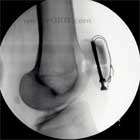
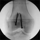
- Displaced, extensor mechanism disrupted, transverse fracture
-Treatment = ORIF with cannulated screws and tension band wire / fiberwire.
- Displaced, extensor mechanism disrupted, inferior pole fracture
-Treatment = partial patellectomy (distal pole) and reattachment of the patellar tendon through drill holes. Must maintain Insall-Salvati ratio around 1.0.
- Displaced periprosthetic patella fracture with intact extensor mechanism. Poor clinical results with ORIF due to patella osteonecrosis. Recommend immobilization in extension either with a long-leg cast or knee immobilizer. (Berry DJ. Patellar fracture following total knee arthroplasty. J Knee Surg. 2003 Oct;16(4):236-41)
Patella Fracture Associated Injuries / Differential Diagnosis
Patella Fracture Complications
Patella Fracture Follow-up Care
- Post-op: knee brace locked in extention, WBAT in brace
- 7-10 Days: Wound check, confirm reduction on xray. Consider allowing knee ROM to some degree based on stability of fixation at surgery.
- 6 Weeks: Evaluate xrays for union. Advance ROM
- 3 Months: progress with ROM and strengthening
- 6 Months: review xrays, return to full activity / sport
- 1Yr: f/u xrays, assess outcome.
Patella Fracture Review References
|


