|


|
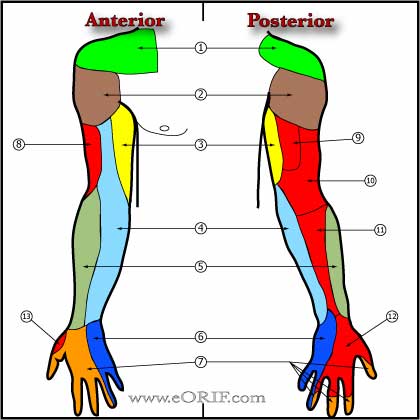
Muscles of the Arm
Muscles of the Forearm
Normal elbow ROM
|
Extension/Flexion
|
0/150
|
|
Pronation
|
80
|
|
Suppination
|
80
|
Flexion from = 0° to 140° +/- 10°
Forearm rotation = 80° to 90° in both supination and pronation.
American Academy of Orthopaedic Surgeons: Joint Motion: Method of Measuring and Recording. Chicago, AAOS, 1965
Boone DC, Azen SP: Normal range of motion of joints in male subjects. JBJS 61A: 756–759, 1979
Morrey BF, Chao EY: Passive motion of the elbow joint. J Bone Joint Surg 58A: 501–508, 1976
Functional elbow ROM (Morrey BF, JBJS 63A;872:1981)
|
Extension/Flexion
|
30/130
|
|
Pronation
|
50
|
|
Suppination
|
50
|
|
|

|
Medial Collateral Ligament
- synonyms: Ulnar collateral ligament.
- consists of anterior bundle, posterior bundle and transverse segment
- Anterior bundle: origin=inferior aspect of the medial epicondyle. Insertion=sublime tubercle of ulna. Primary restraint to valgus stress at the elbow from 30° to 120°. Taut in extension. (Morrey BF, CORR 1991;265:187). Subjected to near-failure tensile stresses during the acceleration phase of the throwing motion.
- Posterior bundle; Origin=medial epicondyle slightly posterior to its most inferior portion. Insertion=broadly onto the olecranon. Taut in flexion.
- MCL is taut in pronation. LCL is taut in supination.
- Morrey BF, CORR 1985;201:84
- Morrey BF, CORR 1991;265:187
- Morrey BF, AJSM 1983;11:315
|
 |
Lateral Collateral Ligament
- Synonyms: lateral ulnar collateral ligament, LUCL, LCL (sometimes considered to include: radial collateral ligament(radial band), annular ligament, & lateral ulnar collateral ligament(ulnar band). Ulnar band of LCL is the primary lateral stabilizer.)
- Origin: lateral epicondyle
- Insertion: tubercle of the supinator crest of the ulna.
- Stabilizer against posterolateral rotational instability
- Taut throughout entire range of motion of the elbow.
- MCL is taut in pronation. LCL is taut in supination.
|
| |
Annular Ligament
- the primary restraint to posterolateral rotatory instability of the elbow is the combination of the lateral collateral and annular ligaments. The principal secondary restraints are the extensor muscles with their fascial bands and intermuscular septa. (Cohen MS, JBJS 1997;79A:225)
|
| |
Leash of Henry
- series of veins encountered in volar approaches to the proximal forearm (Henry approach).
|
| |
Radial Nerve
- in average male the radial nerve at the spiral groove is @15cm from the distal humeral articular surface (Uhl RL, J Orthop Trauma 10:338;1996)
- Proximal to elbow radial nerve is bewteen the brachioradialis and brachialis. Divides into superficial radial nerve and the PIN at the level of the radial head.
|
| |
|
| |
Lateral Antebrachial Cutaneous Nerve
- lateral antebrachial cutaneous nerve-at risk during lateral approaches and anterior approaches for distal biceps tendon repair. Pierces brachial fascia 3cm proximal to the lateral epicondyle and passes 4.5cm medial to the lateral epicondyle. Passes lateral to the biceps tendon. Ant/ post branches supply anteriolateral/posterolateral surfaces of forearm.
|
| |
Medial Antebrachial Cutaneous Nerve
- medial antebrachial cutaneous nerve-at risk during medial approaches. Posterior branch divides into 2-3 branches that cross from 6cm proximal to 6cm distal to medial epicondyle.Lie just superficial to deep fascia
- Painful neuormas / paresthesias can develop due to injury during medial approaches / cubital tunnel surgery (Dellon AL, J Hand Surg 1985;10Br:33).
|
| |
Posterior Interosseus Nerve
- Pierces the supinator muscle and wraps around the radius approximately 1-1.5cm distal to the radial tuberosity.
- The forearm should be fully pronated during lateral approaches to the elbow and fully supinated (moves PIN laterally) during anterior approaches to decrease risk of PIN injury.
- Exposure past the midline of the radial tuberosity during lateral approaches risks PIN injury. (Tornetta P, 3rd, CORR 1997;215)
|
| |
Carrying Angle
- 11° of valgus in men
- 13° of valgus in women
- Beals RK: The normal carrying angle of the elbow. A radiographic study of 422 patients. Clin Orthop 119: 194–196, 1976
- Throwing athletes: may be greater than 15° due to adaptive changes from repetitive stress. (King JW, Clin Orthop 1969;67: 116–123)
- The carrying angle progresses into a more varus alignment as the elbow is flexed (Morrey BF, JBJS 1976;58A:501)
- Carrying angle is difficult to assess if there is any flexion contracture.
|
|

|
Arthroscopy |
| |
Elbow Approaches
References
- Patterson, Surgical Exposures of the elbow. CORR 370:19;2000
- Bryan Morrey Triceps sparing approach CORR 166:188;1982
|
 |
Posterior Utilitarian Approach
- Longitudinal midline posterior incision minimizes risk to cutaneous nerves.
|
 |
Medial Approach to Ulnar Nerve |
 |
|
 |
|
| |
|