|


|
synonyms: DDH, congenital hip dislocation
DDH ICD-10
DDH ICD-9
- 754
- 754.30 unilateral dislocation
- 754.31 bilateral dislocation
- 754.32 unilateral subluxation
- 754.33 bilateral subluxation
- 754.35 dislocation of one hip, subluxation of the other
DDH Etiology / Epidemiology / Natural History
- 1/1000 live births, left hip most common
- More common in children of central European, Native Americans, Laplanders, and native Alaskan descents.
- Etiology: multifactorial, genetic, intruterine mechanical environment,
- Risk of DDH without family history = 0.2%
- Risk of DDH with a parent with DDH = 12%. First-born children are affected twice as often as subsequent siblings. Female infants have histest risk of DDH (@800% of affected infants are female).
DDH Risk factors (five f's)
- first born
- female
- family history
- feet( breech position)
- fluid(oligohydramnios)
DDH Anatomy
DDH Clinical Evaluation
- Ortolani=out-reduces
- Barlow=in-dislocates
- Asymmetric gluteal folds
- Galeazzi sign: apparent femoral length idscrepancy when the legs are held together with the hips and knees flexed.
- Decreased hip abduction
- Amubulatory Patient: flexion contracture, gluteus medius lurch, toe walking, increased lordosis if bilateral
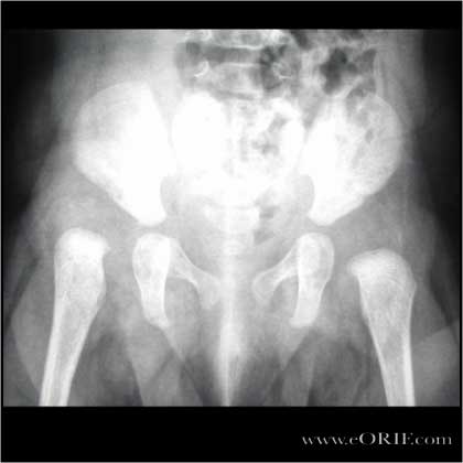
DDH Xray / Diagnositc Tests
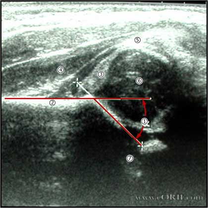
- U/S=gold standard <4months, want Graf(alpha) angle >55 degrees, 50% or more head coverage. (Harcke HT, JBJS 1991;73A:622).
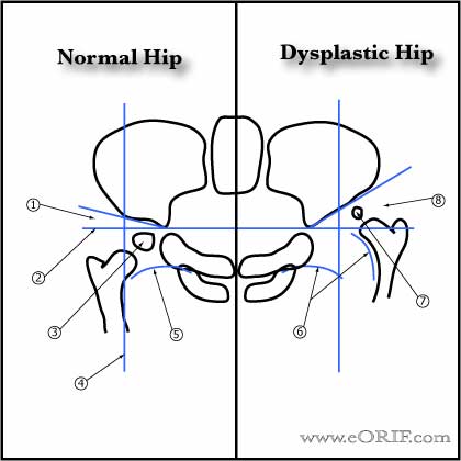
- Patients will likely have severe arthritis if at maturity the lateral center edge angle is <16 degrees and the femoral head is uncovered >1/3.
DDH Classification / Treatment
- Initial treatment for infants =Pavlic harness with weekly U/S f/u until hip is reduced. Pavlic holds hip in flexion(100°) and abduction(limit adduction to neutral).If unreduced in 3-4wks>CR,arthrogram,spica+/-traction,+/-adductor tenotomy. Post-op CT. Cast changes Q6wks follow by night splinting vs Rhino brace
- OR=fails CR, or >18months at presentation. Generally ant approach. Femoral shortening necessary if >24months.
- acetabular index should be 22 degrees at 22 months
- medial approach=can not do capsulorhaphy, higher risk of AVN(25%), <1y/o. Invterval between pectineus and iliopsoas or between adductor brevis and magnus. Risks=medial femoral circumflex vessels
- anteriorleateral approach(Smith-Peterson)=allows capsuloraphy, bikini incision
- Irreducible hip dislocation, moderate dysplasia, moderate subluxation withou complete obliteration of the joint space, preop center-edge angle >10°: Chiari pelvic osteotomy (Ito H, JBJS 2004;86A:1439)..
DDH Associated Anomalies
- Metatarsus adductus
- Hyperextended knees / congential knee dislocation
- Torticollus
DDH Complications
- Pavlik harness complications=femoral nerve palsy, AVN, post acetab wear, medial knee instability, brachial plexus palsy
- Osteonecrosis
- Persistent Acetabular dysplasia
DDH Follow-up Care
- Frequent follow-up with repeat ultrasound/CT indicated to ensure maintenance of reduction.
DDH Review References
- Guille JT, JAAOS 2000;8:232
- Vitale MG, JAAOS 2001;9:401
- Weinstein SL, JBJS 1979;61A:119
- Hayes RJ, ICL 2001;50:535
- Nemeth BA, Narotam V. Developmental dysplasia of the hip. Pediatr Rev. 2012 Dec;33(12):553-61. doi: 10.1542/pir.33-12-553. Review.
- Lee CB, Mata-Fink A, Millis MB, Kim YJ. Demographic differences in adolescent-diagnosed and adult-diagnosed acetabular dysplasia compared with infantile developmental dysplasia of the hip. J Pediatr Orthop. 2013 Mar;33(2):107-11
- °
|