 |
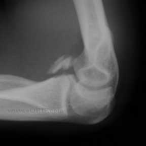
Reagan and Morrey Type III coronoid fracture sustained in a slip and fall. No radiographic evidence of dislocation. Lateral view at presentation 1week after injury shown |
 |
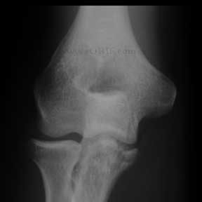
A/P injury view.
Examination revealed postive lateral pivot shift sign and posterolateral rotary drawer test indicative of posterolateral rotary instability.
|
 |
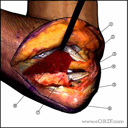
Posteromedial exposure diagram is shown.
- Incision from 6cm proximal to olecranon to 6cm distal to olecranon. Curved around medial border of olecranon to avoid painful scar.
- Medial and lateral skin flaps raised.
- Ulnar nerve identified and transposed anterior to the medial epicondyle.
- Flexor carpi ulnaris incised in line with its origin, leaving a fascial cuff for later repair. FCU subperiosteally elevated exposing anterior band of medial collateral ligament and the coronoid.
- Common Flexor orgin
- Transposed ulnar nerve
- Medial epicondyle
- Anterior band of radial collateral ligament
- Triceps tendon
- Olecranon
- Coronoid
- Reflected FCU origin
|
 |
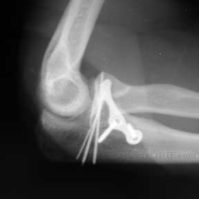
The fracture reduced and fixed with 0.062 k-wires and buttress with a Mayo Clinic Congruent Coronoid plate.
Post-operative lateral view.
|


