|


|
synonyms: colar bone fracture
Clavicle Fx ICD-10
A - Initial encounter for closed fracture
B - initial encounter for open fracture
D - subsequent encounter for fracture with routine healing
G - subsequent encounter for fracture with delayed healing
K - subsequent encounter for fracture with nonunion
P - subsequent encounter for fracture with malunion
S - sequela
Clavicle Fx ICD-9
- 810.01 (closed fracture of sternal end clavicle)
- 810.02 (closed fracture of shaft clavicle)
- 810.03 (closed fracture of acromial end clavicle)
- 810.11 (open fracture of sternal end clavicle)
- 810.12 (open fracture of shaft clavicle)
- 810.13 (open fracture of acromial end clavicle)
Clavicle Fx Etiology / Epidemiology / Natural History
Clavicle Fx Anatomy
- S shaped: anterolateral surface is concave, convex on anteromedial surface.
- First bone to ossify, 5th gestational week
- Only long bone to ossify be intramembranous ossification
- Medial physis=80% of longitudinal growth
- Both acromial and sternal physes can remain open into 3rd decade of life
- Middle 1/3 fractures typically heal with the lateral shaft fragment displaced posterior to the medial shaft fragment / anterior-posterior angulation. (Edleson JG, JSES 2003;12:173).
- Diameter of medullary canal is approximately 7mm at its narrowest point. (Andermahr J, Clin Anat 2006;20:48).
- See also Shoulder Anatomy.
Clavicle Fx Clinical Evaluation
- Pain over clavicle, crepitus and motion at fracture site. Often obvious deformity.
- Evaluate skin over fracture site for tenting, inpending skin compromise
- Ptosis of affected shoulder.
- NV exam of upper extremity indicated; rule out axillary artery/vein injury.
- Auscultate chest to rule out associated pneumothorax.
- Medial clavicle fractures may have dysphagia and shortness of breath.
Clavicle Fx Xray / Diagnositc Tests
- A/P view of clavicle and 30°-45º cephalic tilt view of clavicle.
- Apical oblique (Grashe with 20 cephalad)
- Abduction Lordotic (after ORIF)
- Serendipity view (helps with A/P displacement)
- Axillary
- Zanca 15 degree apical oblique (AC joint)
- Consider chest xray (including both clavicles) if there is significant deformity (shortening) or if concerned for pneumothorax or rib fx's.
- CT with 3-D reconstruction is helpful for segmental fractures or for medial clavicle fractures which may be missed on Chest xrays.
Clavicle Fx Classification / Treatment
- Edinburg Classification (Robinson CM, JBJS 1998;80Br:476)
- Allman Classification (Allman FL, JBJS 49A;774:1967).
- Group I - middle 1/3=69%-85%
-69%-85% of clavicle fractures.
-0.13%-13% non-op nonunion rate overall
-Non-displaced: activity modifications, sling. No difference between sling and figure-8 (Stanly, Injury 19;162:1988), (Andersen K, Acta Orthop Scan 1987;58:71). Figure-8 has been associated with increased axillary pressure sores, compression of the neurovascular bundle, nonunion and patient discomfort. Closed reduction not helpful (Hill, JBJS 1997;79B:537).
-Displaced: Clavicle Fracture ORIF 23515. Displaced = >100% displacement, Shortening >18 mm in males or >14 mm in females (Lazarides S, JSES 2006;15:191). Displaced fractures have improved outcomes with ORIF, decreased nonunion rate, shorter time to union, better shoulder funtinoal scroes (Guerra, Enrico, AJSM2019)(Canadian Orthopaedic Trauma Society JBJS;89:1), (Zlowodzki M, JOT. 2005 Aug;19:504) (Robinson CM, JBJS-A 2013;95:1576). Patients treated nonoperatively are more likely to be dissasisfied with shoulder droop, local bump at fracture site, shoulder asymmetry. Patients treated with surgery at more likey to be disatisfied with local numbness. Robinson CM, JBJS-A 2013;95:1576.
-Operative indications=open fx, NVI, displaced fx with impending skin compromise, shortening >20mm, significant comminution, scapulothoracic dissociation. Consider for neurologic disorders, floating shoulder, bilateral fx, cosmesis, ipsiliateral UE fx, heavy laborers, high level athletes, obvious scapular winging.
-Pediatric: Children younger than 11y/o have excellent outcomes with nonoperative treatment even for displaced fractures. Consider treating children at 12 and older as adults based on displacement.
-See Clavicle Fracture ORIF 23515.
-
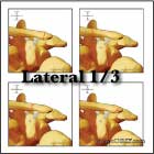
- Group II-lateral 1/3=12%-28%: See Distal Clavicle Fracture 810.03.
- Group III - medial 1/3=3%-6%: generally associated with multisystem trauma; 20% mortality second to associated trauma; generally non-operative treatment (Throckmorton T, JSES 2007;16:49). See Clavicle Fracture-Medial 810.02.
- See Clavicle Fx ORIF Technique.
Clavicle Fx Associated Injuries / Differential Diagnosis
Clavicle Fx Complications
Nonunion(0.1-15%)=little of no progression of clinical or radiographic healing at a minimum of 16 wks
- Risk factors=inadequate immobilization, displacement(no bony contact), comminution, severity of initial trauma, soft-tissue interposition, refracture, primary ORIF, advancing age, female gender,
- Associated complications: limited shoulder motion(61%), neurologic(35%), thoracic outlet syndrome(17%), arterial ischemia(4%) (Jupiter, JBJS 69A;753:1987).
- Treatment = ORIF +/- bone grafting.
Malunion: principle deformity is anterior angulation.
- 46% do not feel fully recovered, 29% pain with activity at 10yr f/u (Nowak, J JSES 2004;13:479).
- Often associated with mild scapular winging.
- Average 2.9cm of shortening.
- Most patients will have major functional deficits including chronic pain, shoulder weakness and thoracic outlet syndrome.
- Treatment = ORIF. (McKee MD, JBJS 2003;85A:790).
Infection
Neurovascular Injury
Hardware failure (Brouwer KM, JSES 2008 IP)
Sequelae of clavicle fractures: shoulder weakness, fatigue, paresthesias, cosmetic deformity, pain at rest or with activity, asymmetry.
Clavicle Fx Follow-up Care
- Post-op: sling for comfort, no overhead motion. Immediate pendelum ROM exercises.
- 10-14 Days: Wound check, sutures removed. Start PT for gentle ROM exercises. No resistive exercises/activities. Sling as needed for comfort.
- 6 weeks: Xrays, if union is evident begin strengthening and resistive exercises. No contant athletics.
- 3 months: Repeat xrays. If no signs of union, consider bone stimulator, see Nonunion.
- Return to sport: there shoulde be both clinical and radiographic evidence of union. Fracture site should be nontender, full shoulder ROM achieved and strength comparable to contralateral side. Generally avoid contact sports and heaving lifting for 4-6 months.
- Nonoperative outcomes: (Robinson CM, JBJS 2004;86A:778).
- Mean time to union = 28.4 weeks for non-operative treatment and 16.4 weeks for operative treatment for displaced clavicle fractures. (McKee MD, JBJS 2007;89A:1).
- Shoulder Outcome measures.
Clavicle Fx Review References
- Zlowodzki M, Zelle BA, Cole PA, Jeray K, McKee MD; Evidence-Based Orthopaedic Trauma Working Group. Treatment of acute midshaft clavicle fractures: systematic review of 2144 fractures: on behalf of the Evidence-Based Orthopaedic Trauma Working Group. J Orthop Trauma. 2005 Aug;19(7):504-7
- Kashif Khan LA, JBJS 2009;91A:447
- Kim W, Orthop Clin N Am 2008;39:491
- Master's Techniques: Shoulder
- Rockwood and Green's Fractures in Adults 6th ed, 2006
- Orthopaedic Knowledge Update: Shoulder and Elbow, No. 3
- Zenni EJ Jr, Krieg JK, Rosen MJ: Open reduction and internal fixation of clavicular fractures. J Bone Joint Surg 1981;63A:147-151.
- Nordqvist A, Redlund-Johnell I, von Scheele A, et al: Shortening of the clavicle after fracture: Incidence and clinical significance. A 5-year follow-up of 85 patients. Acta Orthop Scand 1997;68:349-351.°
- Kim W, McKee MD. Management of acute clavicle fractures. Orthop Clin North Am. 2008 Oct;39(4):491-505
- Canadian Orthopaedic Trauma Society. Nonoperative treatment compared with plate fixation of displaced midshaft clavicular fractures. A multicenter, randomized clinical trial. J Bone Joint Surg Am. 2007 Jan;89(1):1-10.
- McKee MD, Pedersen EM, Jones C, Stephen DJ, Kreder HJ, Schemitsch EH, Wild LM, Potter J. Deficits following nonoperative treatment of displaced midshaft clavicular fractures. J Bone Joint Surg Am. 2006 Jan;88(1):35-40.
- Robinson CM, JBJS-A 2013;95:1576
- MCKEE, MICHAEL D. JBJS: January 2006 - Volume 88 - Issue 1 - p 35–40
|