Proximal Humerus ORIF
synonyms:
Proximal Humerus ORIF CPT
- 23615 Open treatment of proximal humeral fractrue, includes internal fixation, when performed, includes repair of tuberosities.
Proximal Humerus ORIF Anatomy
- Greater tuberosity = insertion of supraspinatus, infraspinatus, and teres minor tendons
- Lesser tuberosity = insertion of subscapularis tendon.
- Displaced greater tuberosity fx is pathognomonic of a longitudinal tear in the rotator cuff at the rotator interval between the supraspinatus and subscapularis tendons.
- Primary blood supply to humeral head is the ascending (arcuate) branch of anterior humeral circumflex artery which runs in the bicipital groove.(Jaberg, JBJS 74A:508;1992) Less significant supplies include the posterior humeral circumflex artery and small vessels enteriing through the rotator cuff insertions.
- Humeral head vascularity after fracture can be estimated by the amount of metaphyseal head extension, <8mm is associated with ischemia; Medial hinge disruption >2mm is associated with ischemia. If both indicate ischmia the positive predictive value of ischemia for an anatomic neck fx is 97%. (Hertel R, JSES 2004;13:427).
- Anatomic Landmarks
-Mean distance between the pectoralis major tendon and the top of the humeral articular surface is 5.6cm (Murachaovsky J, JSES 2006;15:675).
-Normal distance from the greater tuberosity to the superior protion of the articular surface of the humeral head = 7-8mm (Iannotti JP, JBJS 1992;74A:491), (Takase K, JSES 2002;11:557).
-The neck-shaft inclination angle averages 145 degrees. The humeral head is retroveted an average of 30 degrees.
- Most common site of injury to the axillary artery is in the third part(named in relation to the pec minor) of the artery at the origin of the anterior and posterior humeral circumflex arteries.
- Deforming forces: Pectoralis major pulls the shaft medially, anteriorly and internally rotates. Supraspinatus abducts the head fragment in two part fractures. If greater tuberosity is fractured it is pulled superiorly and posteriorly by the suprspinatus and infraspinatus. Lesser tuberosity fractures are pulled medially.
Proximal Humerus ORIF Indications
- 2-, 3-, 4-part proximal humerus fractures
- Valgus-impacted proximal humerus fracture
- Relatively young active patients with good bone density
-
Proximal Humerus ORIF Contraindications
- Poor bone quality
- Medically unstable patient
- Active infection
Proximal Humerus ORIF Alternatives
Proximal Humerus ORIF Pre-op Planning / Case Card
- Develop preoperative plan based on pre-operative radiographs using AO technique.
- Consider getting xrays of normal side to aid in pre-op planning.
- Consider CT scan.
- Anatomic Landmarks
-Mean distance between the pectoralis major tendon and the top of the humeral articular surface is 5.6cm (Murachaovsky J, JSES 2006;15:675).
-Normal distance from the greater tuberosity to the superior protion of the articular surface of the humeral head = 7-8mm (Iannotti JP, JBJS 1992;74A:491), (Takase K, JSES 2002;11:557).
-The neck-shaft inclination angle averages 145 degrees. The humeral head is retroveted an average of 30 degrees.
- Consider augmenting with calcium phosphate cement (Synthes Norian SRS) (Kwon BK, JBJS 2002;84A:951).
- If there are concerns about the patients bone quality, or fracture pattern a hemiarthroplasty prosthesis should be available and the patient should be consented for both ORIF and hemiarthroplasty.
- ORIF Proximal Humerus case card.
Proximal Humerus ORIF Technique
- Pre-operative antibiotics, +/- interscalene block
- General endotracheal anesthesia
- Modified beach-chair position. All bony prominences well padded.
- Examination under anesthesia of affected shoulder.
- Prep and drape in standard sterile fashion. Have a well-padded height adjustable Mayo stand or shoulder positioner available to hold the arm during the case.
- Deltopectoral incision from just medial to AC joint to just lateral to the proximal edge of the biceps muscle belly.
- Identify deltopectoral interval (interval can be found by palpating medial edge of deltoid insertion into clavicle or finding fat layer in interval surrounding cephalic vein.)
- Preserve cephalic vein by ligating any branches to deltoid and taking the cephalic vein and its surrounding tissues medially.
- Incise clavipectoral fascia adjacent to the conjoined tendon up to the coracoacromial ligament.
- Release upper 1/3 of pectoralis tendon if needed for exposure.
- Ensure the anterior humeral circumflex vessels are protected and preserved.
- Identify the long head of the biceps tendon and ensure that it is preserved thoughtout the case.
- Identify the fracture fragments. The key to identifying the various components is the long head of the biceps tendon. The lesser tuberosity and subacapularis tendon are medial to the long head tendon. The greater tuberosity and supraspinatus are lateral. Generally splinting the rotator interval between the tuberosities provides adequate exposure to the proximal humerus.
- Mobilize the tuberosity fragments. Tag them with suture as needed.
- Gently identify the humeral head fragment, being careful to avoid any neurovascular injury. Confirm that the head fragment is not split or impacted and the cartilage is intact.
- Reduce that fragments into anatomic position. The humeral head can usually be reduced by externally rotation the arm and gentle pushing and rotating the head into its anatomic position.
- The fragments are then anatomically reduced and temporarily fixed using k-wires or suture. Placing a non-absorbable #5 suture in a figure-8 fashion is often beneficial to maintain the reduction during plate placement and also serves additional fixation.
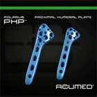
- Place a proximal humeral plate as selected in the preoperative plan using AO technique and as instructed in the manuctures technique guide.
- Pack allograft bone chips / demineralized bone graft as needed to improve healing.
- Repair the rotator interval.
- Irrigate.
- Close in layers.
Proximal Humerus ORIF Complications
- Primary / secondary screw perforation of the humeral head (SudKamp N, JBJS 2009;91A:1320).
- Subacromial impingement
- Nerve injury palsy: Axillary
- Infection
- Loss of reduction
- Need for further surgery
- Hardware failure
- Stiffness
- Pain
- Avascular necrosis of humeral head
- Nonunion
- DVT/PE
- Risks of Anesthesia including heart attack, stroke and death.
- Risks of surgery include but are not limited to: Primary / secondary screw perforation of the humeral head, Subacromial impingement, Nerve injury palsy: Axillary, Infection, Loss of reduction, Need for further surgery, Hardware failure, Stiffness, Pain, Avascular necrosis of humeral head, Nonunion ,DVT/PE, Risks of Anesthesia including heart attack, stroke and death.
Proximal Humerus ORIF Follow-up
- Patients are placed in a shoulder immobilzer with an abduction pillow (Ultrasling) post-operatively. Pendulum, elbow, wrist, hand ROM is started immediately.
- F/U at 7-10 days to remove sutures, check xrays and start passive ROM in physical therapy.
- Active ROM and strengthening are started after xray evidence of fracture healing.
- Return of ROM and strength can take 6months to 1 year.
- Proximal Humerus Fracture Rehab Protocol.
- Shoulder outcome measures.
Proximal Humerus ORIF Outcomes
Proximal Humerus ORIF References
|
|


